Volume: 3 Issue: 3
Large Soft Tissue Masses of the Thigh and Groin That Mimic or Hide Malignancy: A Case Series of a Giant Schwannoma, a Chronic Expanding Haematoma and Metastatic Melanoma
Year: 2026, Page: 141-144, Doi: https://doi.org/10.71325/ajjms.v3i3.26.57
Received: Oct. 7, 2026 Accepted: Oct. 22, 2026 Published: Oct. 31, 2026
Abstract
Large soft tissue masses of the thigh and groin can look like sarcoma when they are benign, and a malignant mass can present away from its primary site. We describe three such masses: a giant intermuscular schwannoma (8.8 cm) appearing on MRI as a well-encapsulated, T2-hyperintense lesion; a chronic expanding haematoma (10 cm) presenting 8 years after blunt trauma with heterogeneous signals and fluid-fluid levels on MRI; and a metastatic malignant melanoma presenting as a 12 cm hard inguinal mass from a "hidden" primary in the toe, previously excised as a "corn". All three patients underwent surgery, with no recurrence at 1 and 3 months. Size was an unreliable predictor of malignancy and misled in both directions; these cases support interpreting MRI alongside the history and examining the distal extremity in any inguinal or thigh mass.
Keywords: Schwannoma, Chronic expanding haematoma, Melanoma, Soft tissue mass, Sarcoma mimic
INTRODUCTION
Large soft tissue masses of the thigh and groin often present a diagnostic dilemma, because size alone does not separate benign lesions from malignant ones. The thigh can harbour lesions that reach "giant" proportions, defined here as exceeding 5 cm in greatest dimension, before becoming symptomatic[1, 2]. Although schwannomas are common benign peripheral nerve sheath tumours, they typically present as small, solitary lesions; giant variants in the extremities are unusual and require sophisticated imaging to differentiate from sarcomas[1, 3]. Chronic expanding haematomas can also mimic aggressive soft tissue sarcomas on imaging[4]. Conversely, a hard inguinal mass may be a secondary deposit from a neglected primary site, such as a subungual or plantar malignant melanoma. Accurate diagnosis needs examination, MRI and histopathology together.
We describe a giant schwannoma, a chronic expanding haematoma and a metastatic melanoma, to show how benign lesions can look like sarcoma and how a malignancy can be missed when the primary lesion is elsewhere.
MATERIALS AND METHODS
These three patients were retrospectively reviewed from affiliated tertiary care teaching hospitals in Southern India (January 2020 to December 2022). A retrospective series was chosen because larger prospective studies are difficult for rare pathologies. They are part of a six-patient cohort; the other three patients are reported separately. Inclusion criteria were a giant (>5 cm) or rare soft tissue mass of the thigh or groin, a challenging preoperative diagnosis or one that conflicted with the final histopathology, and complete clinical records, high-resolution imaging (USG/MRI) and clinical or intraoperative photographs. Common lesions (simple lipomas, superficial sebaceous cysts, acute pyogenic abscesses) and cases with incomplete follow-up or no histopathological confirmation were excluded. Of the eligible patients, three were selected because they best illustrated benign lesions that mimic sarcoma and a malignancy presenting away from its primary site. Clinical data and preoperative imaging (ultrasound and/or MRI) were retrieved from institutional records. Diagnostic work-up included fine-needle aspiration cytology (FNAC) or core needle biopsy, as indicated. Surgery was performed under regional or general anaesthesia, with follow-up at 1 and 3 months to assess recurrence or complications. The study was conducted in accordance with the Declaration of Helsinki (1964) and its subsequent amendments, including the 2000 revision, and was approved by the Institutional Ethics Committee (IEC) of the authors' institution (approval details withheld for blinded review), under the project title "Diagnostic Challenges in Large Thigh Masses: A Comparative Case Series of Rare Musculoskeletal Pathologies", of which the present three-case series is a subset. All clinical data and photographs were de-identified, and written informed consent for publication of clinical details and de-identified images was obtained from all participants or their legal guardians.
CASE SERIES
Case 1: Giant Schwannoma of the Upper Thigh
A 56-year-old man presented with an 8.8 cm firm, non-tender, mobile mass in the left anteromedial thigh ([Fig. 1]a). It caused mild paraesthesia in the distal distribution, and its size initially raised suspicion of a soft tissue sarcoma. MRI demonstrated a well-encapsulated lesion, hyperintense on T2-weighted imaging ([Fig. 1]b). To prevent permanent neurological deficit, an "S-shaped" skin incision was used, allowing wider exposure of the nerve of origin (likely a branch of the femoral or obturator nerve) and intracapsular enucleation ([Fig. 1]c). The wound was closed over a drain ([Fig. 1]d), and postoperative recovery was uneventful. The macroscopic specimen was a solid, well-circumscribed 8.8 cm mass ([Fig. 1]e). Histopathology revealed Antoni A (dense cellularity with Verocay bodies) and Antoni B (loose myxoid tissue) areas, confirming a benign schwannoma ([Fig. 1]f). There was no recurrence at 1 and 3 months.
Case 2: Chronic Expanding Haematoma (CEH)
A 42-year-old man presented with a 10 cm swelling that developed 8 years after blunt trauma ([Fig. 2]a); this latent period is a classic feature of CEH. MRI showed heterogeneous signals and fluid-fluid levels ([Fig. 2]b), features that can mimic an undifferentiated pleomorphic sarcoma (formerly malignant fibrous histiocytoma) or synovial sarcoma. Surgical exploration revealed 300 mL of dark "crankcase-oil" fluid and organised clots ([Fig. 2]c). The diagnosis was confirmed on histopathological examination of the excised tissue, and there was no recurrence at 1 and 3 months.
Case 3: Metastatic Malignant Melanoma (The "Hidden" Primary)
A 48-year-old woman presented with a hard inguinal mass measuring 12 cm ([Fig. 3]a). The inguinal mass was the presenting symptom, but the underlying disease was in the foot: examination revealed that the "corn" excised from her big toe a year earlier was actually a primary acral lentiginous melanoma ([Fig. 3]b). The metastatic inguinal deposit was surgically excised, and histopathological examination confirmed metastatic malignant melanoma. There was no recurrence at 1 and 3 months.
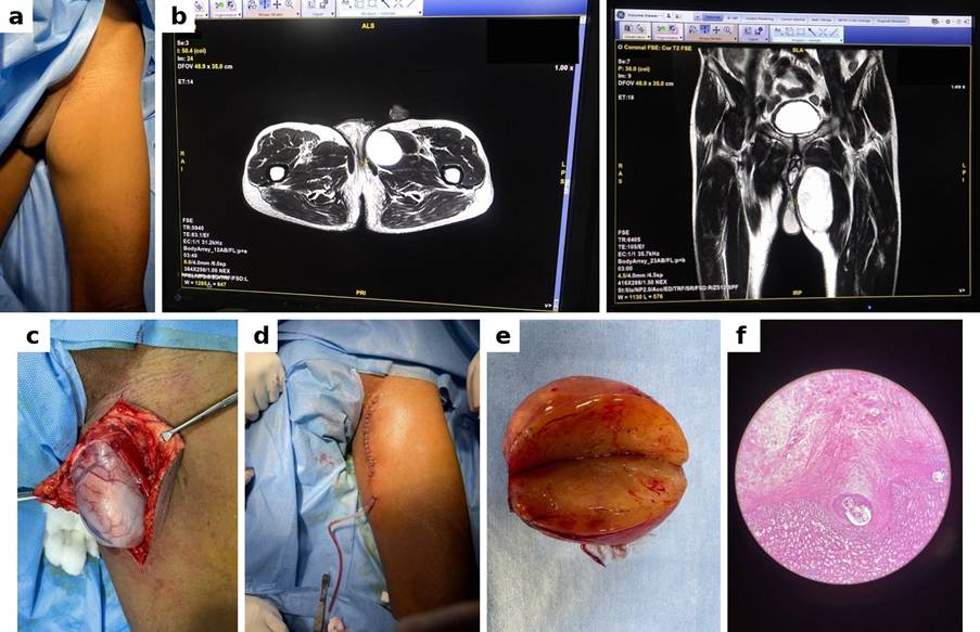
Fig. 1: Giant schwannoma. (a) Pre-operative 8.8 cm thigh mass; (b) T2-weighted MRI, coronal and axial; (c) Intra-operative tumour; (d) Closure with drain; (e) Excised specimen; (f) Photomicrograph showing Antoni A and B areas
DISCUSSION
In these three masses, size was an unreliable guide to malignancy, in both directions. A giant schwannoma (8.8 cm) and a chronic expanding haematoma (10 cm) resembled sarcoma, whereas a 12 cm hard inguinal mass
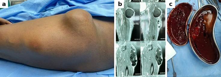
Fig. 2: Chronic expanding haematoma. (a) 10 cm thigh mass; (b) MRI showing heterogeneous signals and fluid–fluid levels within tensor fasciae latae; (c) Evacuated dark “crankcase-oil” fluid and organised clots
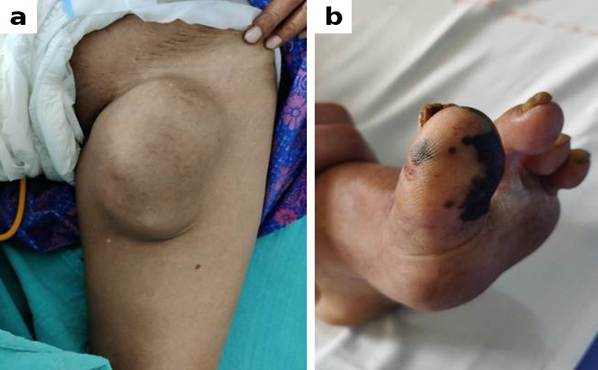
Fig. 3: Metastatic malignant melanoma (the “hidden” primary). (a) Hard, irregular 12 cm metastatic inguinal mass; (b) Primary melanoma of the big toe, with pigmentation at the distal tip and nail region
was a metastasis from an unrecognised primary in the toe.
Clinical size and consistency often fail to distinguish benign mimics from aggressive malignancies, so a combined clinical, radiological and histopathological approach is needed.
Although schwannomas typically measure less than 5 cm, Case 1 reached 8.8 cm. Giant schwannomas are rare and are often misdiagnosed preoperatively as liposarcomas or undifferentiated pleomorphic sarcomas[1, 3]. A well-encapsulated, T2-hyperintense lesion on MRI raises the possibility of a nerve sheath tumour, although MRI alone cannot exclude sarcoma. In our patient, the S-shaped incision allowed intracapsular enucleation with an uneventful recovery[3, 5].
Chronic expanding haematoma in Case 2 mimics malignancy due to its heterogeneous MRI signal, caused by the breakdown of blood products[4]. Unlike simple haematomas, CEHs grow due to the high osmotic pressure of degenerate haemoglobin, which induces recurrent micro-bleeding from a friable pseudocapsule. Surgical removal rather than simple aspiration is therefore generally advised, with excision of the pseudocapsule to prevent recurrence.
Case 3 shows a malignancy that was missed because the primary lesion was elsewhere. A hard inguinal mass may be mistaken for a primary lymphoma or soft tissue sarcoma, so any groin lump requires meticulous examination of the whole foot, including the soles, nail beds and interdigital spaces. Acral melanomas of the sole and nail region are frequently misdiagnosed as benign conditions, including warts and calluses, which delays diagnosis[6].
Limitations
This is a small (n=3) retrospective series from affiliated tertiary care teaching centres, so the findings may reflect local referral patterns and cannot support statistical analysis. Follow-up was short (3 months), which limits comment on recurrence and on the course of the metastatic melanoma. Symptom duration was not documented for two patients, and detailed histopathological and staging data, treatment of the primary toe lesion and any adjuvant therapy for the melanoma are not reported here. The study also relies on MRI, which may not be readily available in resource-limited settings.
CONCLUSION
Size alone is a poor predictor of malignancy in large masses of the thigh and groin. Based on these cases, we suggest that clinicians: (1) examine the foot carefully in any inguinal mass to exclude an occult primary malignancy such as acral lentiginous melanoma; (2) interpret MRI alongside a detailed history, including trauma, to differentiate benign mimics from soft tissue sarcomas; and (3) recognise a schwannoma pre-operatively, which allows nerve-sparing enucleation.
DISCLOSURE
Informed consent: Written informed consent was obtained from all participants included in the study for the publication of their clinical data and accompanying images.
Conflicts of interest: The authors declare that they have no competing interests.
Data Availability Statement: The datasets used and/or analysed in the current study are available from the corresponding author upon reasonable request.
Declaration of generative AI and AI-assisted technologies in the manuscript preparation process: During the preparation of this work, the author(s) used Paperpal AI in order to help with language refinement and proofreading. After using this tool/service, the author(s) critically reviewed and edited the content as needed and take(s) full responsibility for the content of the published article.
Funding: This research received no specific grants from any funding agency in the public, commercial, or not-for-profit sectors.
Acknowledgements: We thank all Surgery Department faculty and postgraduates at JSS Medical College and Hospital, Mysuru, for their support in preparing this article.
Author Contributions: ML contributed to the conceptualisation, data collection, manuscript writing, and editing. TN contributed to clinical supervision, data interpretation, and critical review of the manuscript. All authors approved the final version of the manuscript.
References
1. Humphrey PA, Dehner LP. Peripheral nerve tumours. In: Dehner LP, Humphrey PA, Pfeifer JD, editors. The Washington manual of surgical pathology. 2nd ed. Philadelphia: Lippincott Williams & Wilkins; 2012. pp. 771-787
2. Curran RC, Crocker J. Schwannoma. In: Curran's atlas of histopathology. 4th ed. Oxford: Oxford University Press; 2000. pp. 90-91
3. Rhanim A, El Zanati R, Mahfoud M, Berrada MS, El Yaacoubi M. A rare cause of chronic sciatic pain: Schwannoma of the sciatic nerve. Journal of Clinical Orthopaedics and Trauma. 2013; 4 (2). Available from: https://doi.org/10.1016/j.jcot.2013.04.001
4. Liu PT, Leslie KO, Beauchamp CP, Cherian SF. Chronic expanding hematoma of the thigh simulating neoplasm on gadolinium-enhanced MRI. Skeletal Radiology. 2006; 35 (4). Available from: https://doi.org/10.1007/s00256-005-0042-8
5. Knight DM, Birch R, Pringle J. Benign solitary schwannomas. The Journal of Bone and Joint Surgery. British volume. 2007; 89-B (3). Available from: https://doi.org/10.1302/0301-620x.89b3.18123
6. Soon SL, Solomon AR Jr, Papadopoulos D, Murray DR, McAlpine B, Washington CV. Acral lentiginous melanoma mimicking benign disease: The Emory experience. Journal of the American Academy of Dermatology. 2003; 48 (2). Available from: https://doi.org/10.1067/mjd.2003.63
Copyright
© 2026 Published by Laxmi Memorial Education Trust. This is an open-access article under CC BY 4.0 license. (https://creativecommons.org/licenses/by/4.0/)
Cite this article
Manjunath Lokapur, Thulasivasudevaiah Narayan. Large Soft Tissue Masses of the Thigh and Groin That Mimic or Hide
Malignancy: A Case Series of a Giant Schwannoma, a Chronic Expanding Haematoma and Metastatic Melanoma. AJ J Med Sci 2026;3(3):141-144