Volume: 3 Issue: 2
Green-Synthesized Copper Nanoparticles Derived from Fermented Foxtail Millet Polyphenols Exhibit Antibacterial and Antibiofilm Activity Against Ralstonia solanacearum
Year: 2026, Page: 57-61, Doi: https://doi.org/10.71325/ajjms.v3i2.26.16
Received: March 10, 2026 Accepted: May 20, 2026 Published: Aug. 12, 2026
Abstract
Introduction: Ralstonia solanacearum is a destructive soil-borne bacterium responsible for bacterial wilt in several economically important crops, including tomato, potato, banana, and eggplant. The persistence of this pathogen and increasing concern regarding antimicrobial resistance have encouraged the development of environmentally friendly disease management strategies. Materials and Methods: In the present study, copper nanoparticles (CuNPs) were synthesized through a green synthesis approach using polyphenols extracted from fermented foxtail millets. The synthesized nanoparticles were characterized using UV–visible spectroscopy, Fourier transform infrared spectroscopy (FTIR), dynamic light scattering (DLS), and scanning electron microscopy (SEM). Antibacterial activity of the synthesized nanoparticles against R. solanacearum was evaluated using agar well diffusion, minimum inhibitory concentration (MIC), and biofilm inhibition assays with standard protocols. Results: In the broth dilution assay, optical density decreased from 1.56 ± 0.12 to 0.16 ± 0.09 at 400 µg/mL. The nanoparticles also exhibited strong antibiofilm activity, showing approximately 84% inhibition after 15 h of incubation, whereas the polyphenol extract alone exhibited 41% inhibition. Conclusion: These findings indicate that polyphenol-mediated copper nanoparticles possess promising antibacterial properties and may represent a sustainable approach for managing plant pathogens such as R. solanacearum.
Keywords: Ralstonia solanacearum, Copper nanoparticles, Polyphenols, Foxtail millet, Antibiofilm activity
INTRODUCTION
Bacterial wilt caused by R. solanacearum is one of the most devastating plant diseases worldwide, affecting a wide range of crops. The pathogen infects more than 200 plant species, including economically important crops such as tomato, potato, banana, and eggplant, leading to substantial agricultural losses. Once inside the plant, the bacterium colonises the vascular tissues and disrupts water transport, ultimately leading to wilting and plant death[1-3].
The R. solanacearum species complex consists of genetically diverse strains distributed across various geographical regions. During infection, the bacterium produces several virulence factors, including extracellular polysaccharides, cell wall-degrading enzymes, and proteins that facilitate host colonization. Among these factors, extracellular polysaccharide production plays a critical role in blocking xylem vessels and promoting disease progression. Furthermore, motility and biofilm formation are important traits that enable the pathogen to colonize plant tissues and establish persistent infections[4-7].
Nanotechnology has emerged as a promising approach for addressing challenges in agriculture, particularly in the management of plant diseases. Metallic nanoparticles exhibit unique physicochemical properties such as large surface area, enhanced reactivity, and strong antimicrobial activity. Several studies have demonstrated that nanoparticles such as ZnO, Ag, and CuO can effectively inhibit plant pathogenic bacteria and reduce disease incidence[8-14].
Plant-derived bioactive compounds are increasingly explored as environmentally friendly alternatives to synthetic antimicrobials. Among these compounds, polyphenols represent a diverse class of plant secondary metabolites characterized by aromatic rings containing hydroxyl groups. Polyphenols exhibit a wide range of biological activities, including antioxidant, antimicrobial, antiviral, and anti-inflammatory properties. Importantly, polyphenols can also act as reducing and stabilizing agents in the green synthesis of metal nanoparticles[15-21].
Therefore, the present study aimed to synthesize copper nanoparticles using polyphenols extracted from fermented foxtail millets and to evaluate their antibacterial and anti-biofilm activities against R. solanacearum.
MATERIALS AND METHODS
Bacterial Isolation: Soil samples were collected from the rhizosphere of eggplant plants in Tumkur district, Karnataka, India. Serial dilutions were prepared and plated onto Lysogeny Broth agar plates for bacterial isolation following standard phyto-pathological procedures[1, 2]. Colonies were purified and further characterized using molecular methods.
Molecular Characterization: Genomic DNA was extracted using a CTAB-based method. PCR amplification was performed using the primer pair AU759f and AU760r as described previously[2, 3]. The amplified products were sequenced and analysed using BLAST for species identification.
Fermentation of Millets: Foxtail millets were washed and air-dried. The grains were partially cooked at approximately 70°C and transferred into fermentation flasks. Approximately 100 g of cooked millet was inoculated with yeast culture and sucrose and incubated in the dark. Fermentation experiments were conducted for 3, 7, and 14 days following established methods for enhancing polyphenol production[20, 21].
Extraction of Polyphenols: Fermented extracts were filtered and subjected to solvent extraction using chloroform, followed by ethyl acetate. The extract was concentrated using a rotary evaporator and freeze-dried for further analysis.
Determination of Total Polyphenol Content: Total polyphenol content was determined using the Folin–Ciocalteu method with gallic acid as the standard reference[20, 21].
Green Synthesis of Copper Nanoparticles: 50 mL of polyphenol extract was mixed with 50 mL of 5 mM copper sulphate solution and stirred continuously at 37°C for 24–48 hours. Formation of copper nanoparticles was indicated by a colour change from blue to brown. The nanoparticles were collected by centrifugation, washed with deionized water, and dried in a hot air oven at 70°C following green synthesis protocols[15-18].
Characterization of Nanoparticles: UV-visible spectroscopy, FTIR analysis, DLS measurements, and SEM imaging were used to characterize the synthesized nanoparticles[15-18].
Antibacterial Activity: The antibacterial activity of the synthesized nanoparticles was evaluated using agar well diffusion and MIC assays following standard antimicrobial testing procedures[10-12].
Biofilm Inhibition Assay: Biofilm inhibition was assessed using a microtiter plate assay as described previously[22, 23]. Absorbance was measured at 595 nm to determine biofilm formation.
Statistical Analysis: All experiments were performed in triplicate, and results are expressed as mean ± standard deviation. Statistical significance was determined using one-way ANOVA, with p < 0.05 considered statistically significant.
RESULTS
The bacterial isolate showed 96.3% similarity with R. solanacearum strain CP021766.1 according to BLAST analysis ([Fig. 1] & [Fig. 2]).
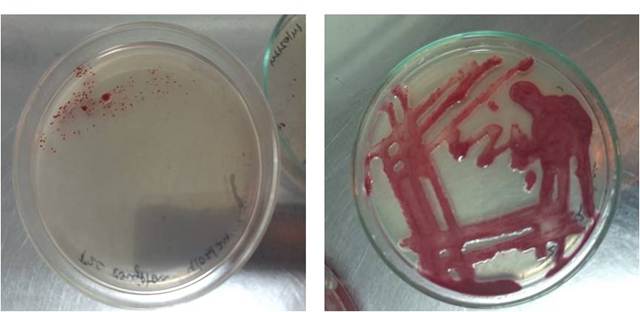
Fig. 1: Images of the plates displaying R. solanacearum isolated from rhizosphere samples
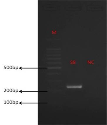
Fig. 2: Agarose gel electrophoresis showing amplified PCR product (~270 bp) confirming molecular identification of R. solanacearum. [M: molecular marker (1 kb ladder); S8: sample; NC: negative control]
Total polyphenol content increased during fermentation and reached the highest value after seven days [Fig. 3].
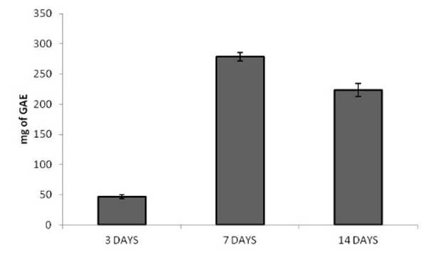
Fig. 3: Histogram showing total polyphenol content of samples obtained from fermented foxtail millets at different incubation times
UV-visible spectroscopy confirmed the formation of copper nanoparticles with a characteristic absorption peak around 240 nm [Fig. 4]. SEM analysis revealed spherical nanoparticles with sizes ranging from 48 to 74 nm ([Fig. 5] & [Fig. 6]).
The agar well diffusion assay demonstrated strong antibacterial activity against R. solanacearum, with a mean inhibition zone of 11.67 ± 0.68 mm [Fig. 7].
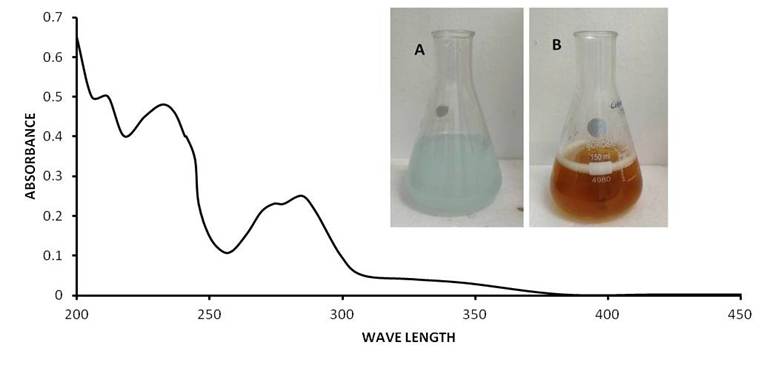
Fig. 4: UV-visible spectra of green synthesized copper nanoparticles
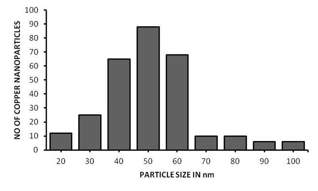
Fig. 5: Histogram showing size distribution of copper nanoparticles obtained from green synthesis
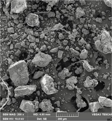
Fig. 6: SEM image presenting spherical morphology of green synthesized copper nanoparticles (48–74 nm)
In the broth dilution assay, optical density decreased from 1.56 ± 0.12 to 0.16 ± 0.09 at 400 µg/mL [Fig. 8].
Biofilm inhibition results showed approximately 84% inhibition after 15 h, whereas the polyphenol extract alone showed 41% inhibition [Fig. 9].
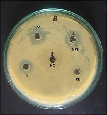
Fig. 7: Agar well diffusion assay showing antibacterial activity. [NC: negative control; PC: positive control; Cu: CuSO₄; E: extract; NPs: synthesized nanoparticles]
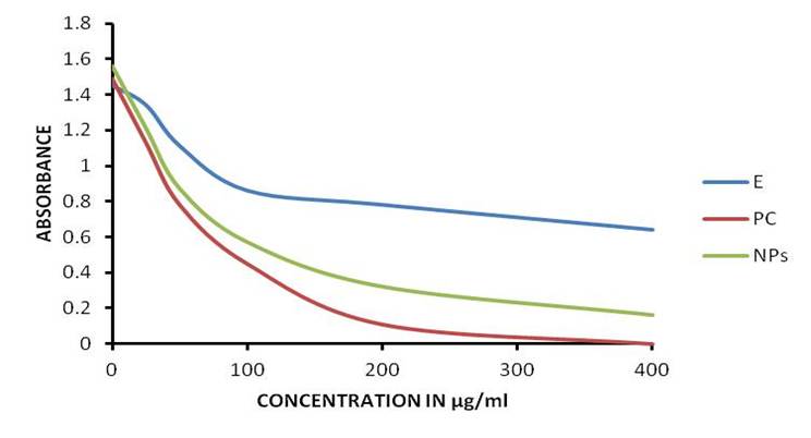
Fig. 8: Broth dilution assay displaying dose-dependent reduction in optical density
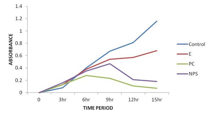
Fig. 9: Biofilm inhibition assay demonstrating reduction in biofilm formation
DISCUSSION
Polyphenols are widely recognized for their antimicrobial activity against various microorganisms. These compounds can disrupt bacterial cell membranes, interfere with enzymatic systems, and affect metabolic processes[20, 21]. In the present study, polyphenols extracted from fermented foxtail millets were successfully used as reducing agents for the synthesis of copper nanoparticles[15-18, 22].
The synthesized nanoparticles demonstrated strong antibacterial activity against R. solanacearum. These nanoparticles interact electrostatically with bacterial cell envelopes, leading to membrane destabilization and increased permeability. In addition, copper nanoparticles generate reactive oxygen species, inducing oxidative stress and damaging cellular macromolecules such as proteins, lipids, and nucleic acids[23-26].
Another mechanism involves the release of copper ions from the nanoparticle surface, which interferes with enzyme activity and cellular respiration. The nanoscale size and high surface-to-volume ratio enhance interaction with bacterial cells, improving antimicrobial efficiency.
Biofilm formation plays a crucial role in the persistence of R. solanacearum. The significant reduction observed suggests that copper nanoparticles interfere with quorum sensing and extracellular polymeric substance production, thereby inhibiting biofilm development[27, 28].
Limitations
This study is limited to in vitro conditions. Further studies should include field-level validation and formulation development.
CONCLUSION
This study demonstrates an environmentally friendly approach for synthesizing copper nanoparticles using polyphenol extracts from fermented foxtail millets. The synthesized nanoparticles showed significant antibacterial and anti-biofilm activity against Ralstonia solanacearum. These findings highlight the potential of green-synthesized nanoparticles as sustainable alternatives for plant disease management.
DISCLOSURE
Conflict of interest: The authors declare no conflicts of interest.
Funding: Nil.
References
1. Hayward AC. Biology and epidemiology of bacterial wilt caused by Ralstonia solanacearum. Annual Review of Phytopathology. 1991; 29 (1). Available from: https://doi.org/10.1146/annurev.py.29.090191.000433
2. Genin S, Denny TP. Pathogenomics of theRalstonia solanacearumSpecies Complex. Annual Review of Phytopathology. 2012; 50 (1). Available from: https://doi.org/10.1146/annurev-phyto-081211-173000
3. Mansfield J, Genin S, Magori S, Citovsky V, Sriariyanum M, Ronald P, et al. Top 10 plant pathogenic bacteria in molecular plant pathology. Molecular Plant Pathology. 2012; 13 (6). Available from: https://doi.org/10.1111/j.1364-3703.2012.00804.x
4. Denny TP. Plant pathogenic Ralstonia species. In: Gnanamanickam SS, editor. Plant-associated bacteria. Dordrecht: Springer. 2006; Available from: https://doi.org/10.1007/978-1-4020-4538-7_16
5. Peeters N, Guidot A, Vailleau F, Valls M, Genin S, Boucher C, et al. Ralstonia solanacearum, a widespread bacterial plant pathogen in the post-genomic era. Molecular Plant Pathology. 2013; 14 (7). Available from: https://doi.org/10.1111/mpp.12038
6. Vasse J, Frey P, Trigalet A. Microscopic studies of intercellular infection and protoxylem invasion of tomato roots by Pseudomonas solanacearum. Molecular Plant-Microbe Interactions. 1995; 8 (2). Available from: https://doi.org/10.1094/mpmi-8-0241
7. Denny TP. Plant pathogenic Ralstonia species virulence factors. Annual Review of Phytopathology. 2006; 44:269–289.
8. Yuliar, Nion YA, Toyota K. Recent trends in control methods for bacterial wilt diseases caused by Ralstonia solanacearum. Microbes and environments. 2015; 30 (1). Available from: https://doi.org/10.1264/jsme2.me14144
9. Agrios GN. Plant pathology. 5th ed. Burlington: Elsevier Academic Press; 2005.
10. Rai M, Yadav A, Gade A. Silver nanoparticles as a new generation of antimicrobials. Biotechnology Advances. 2009; 27 (1). Available from: https://doi.org/10.1016/j.biotechadv.2008.09.002
11. Jo YK, Kim BH, Jung G. Antifungal Activity of Silver Ions and Nanoparticles on Phytopathogenic Fungi. Plant Disease. 2009; 93 (10). Available from: https://doi.org/10.1094/pdis-93-10-1037
12. Mishra S, Singh HB. Biosynthesized nanoparticles as a sustainable tool in plant disease management. Frontiers in Microbiology. 2015; 6:141.
13. Rai M, Ingle AP, Gupta I, Gaikwad S, Gade A, da Silva SS. Nanotechnology based anti-infectives and therapeutics. Journal of Applied Microbiology. 2018;125(5):1259–1273.
14. Khan I, Saeed K, Khan I. Nanoparticles: Properties, applications and toxicities. Arabian Journal of Chemistry. 2019; 12 (7). Available from: https://doi.org/10.1016/j.arabjc.2017.05.011
15. Ahmed S, Ahmad M, Swami BL, Ikram S. A review on plants extract mediated synthesis of silver nanoparticles for antimicrobial applications: A green expertise. Journal of Advanced Research. 2016; 7 (1). Available from: https://doi.org/10.1016/j.jare.2015.02.007
16. Mittal AK, Chisti Y, Banerjee UC. Synthesis of metallic nanoparticles using plant extracts. Biotechnology Advances. 2013; 31 (2). Available from: https://doi.org/10.1016/j.biotechadv.2013.01.003
17. Prasad R, Pandey R, Barman I. Engineering tailored nanoparticles with microbes: quo vadis?. WIREs Nanomedicine and Nanobiotechnology. 2016; 8 (3). Available from: https://doi.org/10.1002/wnan.1363
18. Iravani S. Green synthesis of metal nanoparticles using plants. Green Chemistry. 2011; 13 (10). Available from: https://doi.org/10.1039/c1gc15386b
19. Singh P, Kim YJ, Zhang D, Yang DC. Biological Synthesis of Nanoparticles from Plants and Microorganisms. Trends in Biotechnology. 2016; 34 (7). Available from: https://doi.org/10.1016/j.tibtech.2016.02.006
20. Cushnie TPT, Lamb AJ. Antimicrobial activity of flavonoids. International Journal of Antimicrobial Agents. 2005;26(5):343–356.
21. Daglia M. Polyphenols as antimicrobial agents. Current Opinion in Biotechnology. 2012; 23 (2). Available from: https://doi.org/10.1016/j.copbio.2011.08.007
22. Bhuyan T, Mishra K, Khanuja M, Prasad R, Varma A. Biosynthesis of zinc oxide nanoparticles from Azadirachta indica for antibacterial and photocatalytic applications. Materials Science in Semiconductor Processing. 2015; 32 Available from: https://doi.org/10.1016/j.mssp.2014.12.053
23. Lemire JA, Harrison JJ, Turner RJ. Antimicrobial activity of metals: mechanisms, molecular targets and applications. Nature Reviews Microbiology. 2013; 11 (6). Available from: https://doi.org/10.1038/nrmicro3028
24. Rai M, Yadav A, Gade A. Silver nanoparticles as a new generation of antimicrobials. Biotechnology Advances. 2009; 27 (1). Available from: https://doi.org/10.1016/j.biotechadv.2008.09.002
25. Ahmed S, Ahmad M, Swami BL, Ikram S. Plant extract mediated synthesis of silver nanoparticles: A review. Journal of Advanced Research. 2016; 7 (1). Available from: https://doi.org/10.1016/j.jare.2015.02.007
26. Prasad R, Pandey R, Barman I. Engineering nanoparticles with microbes. WIREs Nanomedicine and Nanobiotechnology. 2016;8(3):316–330.
27. Mishra S, Singh HB. Biosynthesized nanoparticles as sustainable plant disease management tools. Frontiers in Microbiology. 2015; 6:141.
28. Khan I, Saeed K, Khan I. Nanoparticles: Properties, applications and toxicities. Arabian Journal of Chemistry. 2019; 12 (7). Available from: https://doi.org/10.1016/j.arabjc.2017.05.011
Copyright
© 2026 Published by Laxmi Memorial Education Trust. This is an open-access article under CC BY 4.0 license. (https://creativecommons.org/licenses/by/4.0/)
Cite this article
Maithili Acharya, Manjula Shantaram. Green-Synthesized Copper Nanoparticles Derived from Fermented Foxtail Millet Polyphenols Exhibit Antibacterial and Antibiofilm Activity Against Ralstonia solanacearum. AJ J Med Sci 2026;3(2):57-61