Volume: 3 Issue: 3
Correlation Between Infection Severity and Immunological Biomarkers in Patients with Entamoeba histolytica
Year: 2026, Page: 108-114, Doi: https://doi.org/10.71325/ajjms.v3i3.26.33
Received: July 19, 2026 Accepted: Aug. 8, 2026 Published: Sept. 5, 2026
Abstract
Introduction: Amoebiasis is the third most prevalent parasite cause of morbidity and death, especially in nations with unsanitary conditions in Africa, India, and Central and South America. The current study was conducted to evaluate values of immunological parameters, hepcidin, ferritin, hematological indicators in patients with Entamoeba histolytica. Materials and Methods: The study included a total of 90 participants, that divided into 2 groups as 30 control and 60 patients infected with E. histolytica, which sub-divided according to severity into moderate and sever, each of them with 30 patients. The age range for both males and females was between 1 and > 65 years. All participants recruited between 4/1/2025 to 1/10/2025 were taken from people with diarrhea from Ameriyat Al-Sumoud district, Ameriyat General Hospital and outpatient clinics, in Anbar city. Results: The present study showed that IL-6 increased from 4.40 ± 0.82 in healthy individuals to 24.09 ± 5.10 in severe cases, and IL-10 increased from 6.51 ± 1.47 to 22.53 ± 4.50. In contrast, hepcidin levels decreased significantly with increasing infection severity, decreasing from 5.03 ± 1.32 g/dL in the control group to 0.88 ± 0.44 g/dL in severe cases. Hemoglobin levels decreased from 12.03 ± 1.08 g/dL in the control group to 9.20 ± 0.82 g/dL in severe cases. The MCV, MCH, and MCHC values also decreased, while RDW-SD was significantly elevated in patients, particularly in severe cases. Platelet count showed a significant increase with infection severity, rising from 306.9 ± 88.2 to 666.8 ± 112.3 × 10³/µL. Ferritin levels were also reduced in patients compared to control, particularly in severe cases, decreasing from 105.18 ± 55.4 to 7.48 ± 5.1 ng/mL. Conclusion: According to the results of this investigation, hematological parameters, interleukin-6 (IL-6), interleukin-10 (IL-10), and hepcidin are all potential biomarkers for the detection of the E. histolytica parasite.
Keywords: <I>E. histolytica</I>; Immunological biomarkers; Hematological parameters
INTRODUCTION
According to WHO (2020)[1], amoebiasis is the third most prevalent parasite cause of morbidity and death, especially in nations with unsanitary conditions in Africa, India, and Central and South America. Just 10–20% of those infected with E. histolytica exhibit symptoms. Weight loss, cramping in the abdomen, pain, and watery or bloody diarrhea are symptoms of the disease. Fever and right upper quadrant abdominal pain may be signs of hepatic involvement (liver abscess)[2, 3].
The pathogenic protozoan parasite Entamoeba histolytica is a type of disease-causing protozoan. According to a 2018 report by Sauvey et al.[4], this parasite causes approximately 50 million infections and 70,000 deaths worldwide each year. It has two core stages in its life cycle: the trophozoite and the cyst. It can invade the human body via contaminated food and water, first colonizing the intestinal tract, and is capable of further invading the liver, lungs, and brain. Ninety percent of infected individuals are asymptomatic, but the infection can trigger severe conditions such as bloody diarrhea. Relevant clinical conclusions are cited from studies by Jasni et al., (2022) and Jiang et al., (2024)[5, 6].
Entamoeba histolytica is the causative agent of amebiasis, an intestinal parasitic illness. Humans can contract the single-celled protozoan Entamoeba histolytica through ingesting tainted food or water. Loose poops, cramps, and pain in the abdomen are signs of an amoebic infection. However, the majority of amebiasis patients will not have any significant symptoms. In tropical nations with lax public health measures, amoebae infections are prevalent[7]. This study first clarifies that protozoan trophozoites in the colon hematogenously disseminate to the liver via the portal circulation, and trigger hepatocyte injury through three types of mechanisms: parasite-induced cytotoxicity, inflammatory mediators, and host immune responses. This pathogenic pathway is supported by citations of Kumar et al. (2024)[8] and Sellau et al. (2021)[9]. The study further cites Usuda et al. (2022)[10] to note that the heterogeneity of subsequent liver biochemical changes is determined by four core factors.
When a host contracts an infection with Entamoeba histolytica (E. histolytica), it rapidly initiates a multi-dimensional normal immune response. Drawing on the set of preliminary pathogen-blocking defense mechanisms outlined in Uribe-Querol & Rosales (2020)[11] — which include lymphocytes, mucus secreted by the mucosa of the skeletal system, intestinal peristalsis, and the acidic environment of the stomach — this study proposes that cell-mediated immune responses are the core host defense mechanism against this pathogen. The Toll-like receptor (TLR)-2/4 NFKB plays a role in the initial stage of infection through the interaction of the carbohydrate domain of Gal/GalNAc lectin with intestinal epithelial cells. This interaction results in the generation of inflammatory cytokines such as IL-1B, IL-6, IL-8, IL-12, IL-17, IFN-Y, and TNF-α [12, 13]. host tolerance to the invading trophozoites is maintained by many host variables. IL 10 supports mucosal barrier integrity. IL 10 defective mice were significantly sensitive to intestinal invasion in an amebic colitis model[14]. The liver-expressed antimicrobial peptide hepcidin is secreted mainly by hepatocytes near the portal veins (delivering dietary iron) and by Kupffer cells (sensing bacteria and recycling erythrocytes). In addition, a tiny quantity of hepcidin is produced by both adipocytes and macrophages[15]. Hence, present investigation was carried out for evaluation of values of immunological indices, hepcidin, ferritin, hematological indicators in patients with Entamoeba histolytica.
MATERIALS AND METHODS
Sample collection
The study included a total of 90 participants, that were divided into 2 groups as 30 control and 60 patients infected with E. histolytica, which sub-divided according to severity into moderate and sever, each of them with 30 patients. The mean age in the control group was 31.1 ± 20.4 years, while the mean age in the moderate anemia group was 26.8 ± 20.5 years, and the severe anemia group had a mean age of 31.2 ± 19.5 years. All participants were recruited between 4/1/2025 to 1/10/2025 were taken from people with diarrhea from Ameriyat Al-Sumoud district, Ameriyat General Hospital and outpatient clinics, in Anbar city.
Microscopic examination
Stool samples were obtained using a sterile container. Fresh samples were then analyzed under a light microscope at a high magnification of 40X. A small volume (1-3 ml) of feces. 4-6 ml of venous blood samples were collected by venipuncture. The blood samples were quickly transferred into containers containing EDTA for CBC analysis after sampling. For the serum biochemistry study, another part was moved into tubes that did not contain anticoagulant. Serum was extracted from blood samples centrifuged at 5,000 rpm for 5 minutes at room temperature in tubes that did not contain anticoagulant.
Blood and biochemical tests
Sixty people were diagnosed with the parasite, exhibiting indications of infection. Blood was obtained for the execution of hematological and biochemical analyses. This study conducted two types of medical tests. The seven indicators for hematological complete blood count (CBC) analysis were detected using the pocH-100i® fully automatic hematology analyzer manufactured by Sysmex Corporation, based in Kobe, Japan. The biochemical tests covered two parameters: alkaline phosphatase and ferritin.
Assessment of the IL-6, IL-10, and Hepcidin
The concentration of IL-6, IL-10, and Hepcidin was quantified via ELISA. The plate has been pre-treated with a human anti- (IL-6, IL-10, and Hepcidin) antibody. In this experiment, three target molecules IL-6, IL-10, and Hepcidin were first introduced into biological samples. After immune complexes were coated using the corresponding biotinylated human-derived antibody conjugates, the substrate was added, followed by the addition of an acidic stop solution. Absorbance was then detected at a wavelength of 450 nm to complete the quantitative detection of the target proteins.
Assessment of ferritin
Ferritin levels were measured using an automatic biochemistry analyzer within the scope of serum biochemistry.
Statistical analysis
Analyses for this study were completed using IBM SPSS Statistics 26.0, while GraphPad Prism 8 was used for data visualization and result validation. Continuous variables are presented as mean ± standard deviation, and categorical variables are summarized by frequency and percentage (%). Comparisons among the three study groups (controls, moderate anemia, and severe anemia) were performed using one-way analysis of variance (ANOVA) for continuous variables. Upon obtaining statistically significant results, Bonferroni post-hoc test was applied to determine pairwise differences between groups.
Categorical variables, including gender and age group distributions, were analyzed using the Chi-square (χ²) test, with Fisher’s Exact test applied when the expected cell counts were less than 5. Correlation between biomarkers and hematological parameters was assessed using Spearman’s rank correlation coefficient (r), to evaluate the strength and direction of associations. GraphPad Prism was specifically utilized for generating graphs, including bar charts and correlation plots, as well as for visual interpretation of statistical findings. A p-value of less than 0.05 was considered statistically significant.
RESULTS
The total sample of 90 participants was divided equally between the control group and the two groups of parasitic infection depending on symptoms as moderate and severe. Each group comprised 30 individuals, representing 33.3% of the total sample.
A significant increase in IL-6 and IL-10 levels was observed in patients compared to the control group. IL-6 increased from 4.40 ± 0.82 in healthy individuals to 24.09 ± 5.10 in severe cases, and IL-10 increased from 6.51 ± 1.47 to 22.53 ± 4.50. In contrast, hepcidin levels decreased significantly with increasing infection severity, decreasing from 5.03 ± 1.32 g/dL in the control group to 0.88 ± 0.44 g/dL in severe cases. Hb, PCV, and RBC levels decreasing significantly with infection severity. Hemoglobin levels decreased from 12.03 ± 1.08 g/dL in the control group to 9.20 ± 0.82 g/dL in severe cases. The MCV, MCH, and MCHC values also decreased RDW-SD was significantly elevated in patients, particularly in severe cases.
PLT showed a significant increase with infection severity, rising from 306.9 ± 88.2 to 666.8 ± 112.3 × 10³/µL. Ferritin level also reduced in patients compared to control, particularly in severe cases, decreasing from 105.18 ± 55.4 to 7.48 ± 5.1 ng/mL. In contrast, non-significant difference between the groups (P = 0.631), regarding ages as shown in [Table. 1].
The inflammatory cytokines IL-6 and IL-10 showed highly significant differences between all groups (P < 0.001), including the comparison between moderate and severe cases. Hepcidin also showed a significant decrease between all groups, even between moderate and severe cases (P = 0.0002).
| Variable | Controls (mean±SD) | Moderate (mean±SD) | Severe (mean±SD) | p-value | Sig. |
|---|---|---|---|---|---|
| IL-6 | 4.40 ± 0.82 | 17.07 ± 3.42 | 24.09 ± 5.10 | <0.001 | *** |
| IL-10 | 6.51 ± 1.47 | 17.99 ± 4.12 | 22.53 ± 4.50 | <0.001 | *** |
| Hepcidin | 5.03 ± 1.32 | 1.25 ± 0.62 | 0.88 ± 0.44 | <0.001 | *** |
| HGB (g/dL) | 12.03 ± 1.08 | 9.80 ± 0.99 | 9.20 ± 0.82 | <0.001 | *** |
| HCT (%) | 40.50 ± 3.12 | 33.37 ± 2.50 | 31.43 ± 2.30 | <0.001 | *** |
| RBCs (x10⁶/µL) | 4.93 ± 0.48 | 3.70 ± 0.55 | 3.14 ± 0.44 | <0.001 | *** |
| MCV (fL) | 89.78 ± 5.20 | 78.23 ± 4.10 | 71.89 ± 3.80 | <0.001 | *** |
| MCHC (g/dL) | 34.45 ± 1.20 | 30.87 ± 1.40 | 30.38 ± 1.30 | <0.001 | *** |
| MCH (pg) | 29.85 ± 2.10 | 24.40 ± 1.80 | 23.71 ± 1.50 | <0.001 | *** |
| RDW-SD | 46.21 ± 4.20 | 58.13 ± 5.50 | 63.75 ± 6.10 | <0.001 | *** |
| PLT (x10³/µL) | 306.9 ± 88.2 | 528.5 ± 95.4 | 666.8 ± 112.3 | <0.001 | *** |
| Ferritin (ng/mL) | 105.18 ± 55.4 | 10.71 ± 8.2 | 7.48 ± 5.1 | <0.001 | *** |
| Age (years) | 31.12 ± 20.1 | 26.82 ± 18.5 | 31.20 ± 21.4 | 0.631 | ns |
Table 1: Comparison level of immunological, hematological, biochemical between study groups
*** p < 0.001· ** p < 0.01 · * p < 0.05 · ns = not significant
| Variable | Controls vs Moderate | Controls vs Severe | Moderate vs Severe |
|---|---|---|---|
| IL-6 | *** < 0.001 | *** < 0.001 | *** < 0.001 |
| IL-10 | *** < 0.001 | *** < 0.001 | *** < 0.001 |
| Hepcidin | *** < 0.001 | *** < 0.001 | ** p = 0.0002 |
| HGB | *** < 0.001 | *** < 0.001 | ns (p = 0.601) |
| HCT | *** < 0.001 | *** < 0.001 | ns (p = 0.601) |
| RBCs | *** < 0.001 | *** < 0.001 | ** p = 0.003 |
| MCV | *** < 0.001 | *** < 0.001 | *** < 0.001 |
| MCHC | *** < 0.001 | *** < 0.001 | ns (p = 0.569) |
| MCH | *** < 0.001 | *** < 0.001 | ns (p = 0.055) |
| RDW-SD | *** < 0.001 | *** < 0.001 | *** < 0.001 |
| PLT | *** < 0.001 | *** < 0.001 | *** < 0.001 |
| FERRITIN | *** < 0.001 | *** < 0.001 | ns (p = 0.293) |
Table 2: Post-Hoc Pairwise Comparisons (Bonferroni-Corrected) of immunological, hematological, biochemical between study groups
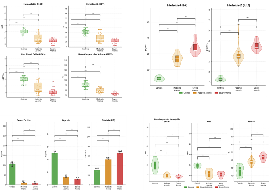
Fig. 1: Post-Hoc Pairwise Comparisons (Bonferroni-Corrected) of immunological, hematological, biochemical between study groups
Regarding Ferritin, Hb and PCV values showed significant differences between the control group and both patient groups, while no significant differences were
found between moderate and severe cases. RBCs also showed a significant difference between moderate and severe cases. MCV showed highly significant differences between all groups. In contrast, MCHC and MCH values did not show significant differences between moderate and severe cases. RDW-SD also showed highly significant differences between all groups. For PLT highly significant differences were also observed between all groups as shown in [Table. 2] and [Fig. 1].
|
Biomarker pair |
r |
p-value |
Direction |
|---|---|---|---|
|
IL-6 vs IL-10 |
0.839 |
<0.001 |
Positive |
|
IL-6 vs PLT |
0.815 |
<0.001 |
Positive |
|
Ferritin vs MCV |
0.816 |
<0.001 |
Positive |
|
MCV vs RBCs |
0.791 |
<0.001 |
Positive |
|
HGB vs RBCs |
0.748 |
<0.001 |
Positive |
|
HGB vs Ferritin |
0.762 |
<0.001 |
Positive |
|
Hepcidin vs PLT |
-0.798 |
<0.001 |
Negative |
|
IL-6 vs MCV |
-0.797 |
<0.001 |
Negative |
|
IL-6 vs Hepcidin |
-0.765 |
<0.001 |
Negative |
|
IL-10 vs Hepcidin |
-0.764 |
<0.001 |
Negative |
Table 3: Spearman Rank Correlation of immunological, hematological, biochemical between study groups
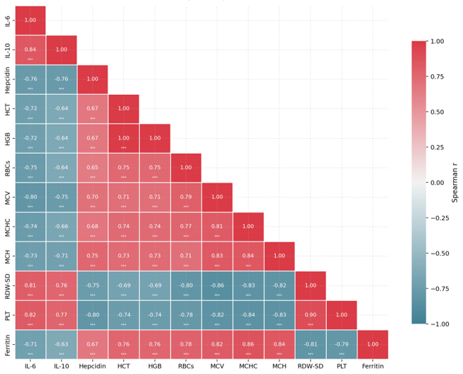
Fig. 2: Spearman Rank Correlation of immuno- logical, hematological, biochemical between study groups
Diverging color scale from r = -1 (blue) to r = +1 (red). *** p < 0.001.
The correlation analysis revealed strong and statistically significant relationships between immunological and hematological markers in patients infected with the parasite. All correlation values were highly significant (P < 0.001), indicating a clear link between inflammation severity, iron imbalance, and hematological changes resulting from the parasitic infection. The results showed a very strong positive correlation between IL-6 and IL-10 (r = 0.839), IL-6 and PLT (r = 0.815), ferritin and MCV (r = 0.816), MCV with RBCs (r = 0.791), HGB and RBCs (r = 0.748), and HGB with ferritin (r = 0.762). In contrast, some variables showed strong inverse relationships. A strong negative correlation was found between Hepcidin and PLT (r = -0.798), IL-6 and MCV (r = -0.797), IL-6 and Hepcidin (r = -0.765), as well as between IL-10 and Hepcidin (r = -0.764) as shown in [Table. 3] and [Fig. 2].
DISCUSSION
The current investigation found that patients with E. histolytica had higher levels of IL-6 and IL-10, which were correlated with severity. This study supports the findings of[16], which demonstrated elevated IL-6 in E. histolytica patients. When compared to healthy controls, individuals with amebic liver abscesses had much greater levels of the cytokine IL-6[17]. Through IL-1β and STAT3, IL-6 contributes to the liver's synthesis of the acute phase reactant C-reactive protein (CRP)[18]. Additionally, IL-6 is a peripheral indicator of acute inflammation in a variety of clinical conditions[17]. It was most recently demonstrated that E. histolytica has a humanlike macrophage migration inhibitory factor (EhMIF) that stimulates IL-6 synthesis, which may further contribute to IL-6 production during invasive amebiasis[19]. IL-6 deficiency enhanced the risk of amebic liver abscess in a mouse model of amebiasis, indicating an important function for IL-6 in the inflammatory processes caused by E. histolytica[20].
This finding is in agreement with that of Ngobeni et al. (2022)[21], who discovered that malnutrition reduces the production of cytokines and the proper response of lymphocytes to these signals. When E. histolytica invades liver tissue, the immune system goes into anti-inflammatory mode. IL-10 has the ability to block the production of proinflammatory cytokines by cells such regulatory T-cells and macrophages, including IFN-γ, IL-2, IL3, TNF-α, and GM-CSF. In addition to its powerful antigen-presenting cell suppression capabilities, it acts as a stimulant to specific T cells (Th2) and mast cells, promotes B cell maturation and antibody production, and more[22].
Due to the high density of this protozoan, nutrients needed to produce blood components are mal-absorbed[23]. These results are consistent with those of Ahmed and Al-Naqeeb (2026)[24], who discovered that people with specific intestinal parasite infections frequently experienced anemia. These results are consistent with those of Ahmed and Al-Naqeeb (2026)[24], who discovered that people with specific intestinal parasite infections frequently experienced anemia. Ferritin is considered an early and highly specific marker since serum ferritin is a measure of body iron reserves and is used to detect iron insufficiency in the absence of concomitant diseases. Children infected with E. histolytica likely have low ferritin levels because the parasite's trophozoite can use ferritin as an iron source[25]. It is known that the intestinal parasite Entamoeba histolytica requires iron for its metabolic processes. As a result, it makes use of the iron present in the liver and the lining of the large intestine, which is where the parasite first appears. According to research by Lopez-Soto et al. (2009)[25], E. histolytica uses ferritin as a crucial iron source during its trophozoite stage by attaching to it via certain proteins. The iron reserves of those infected with this parasite are significantly reduced as a result of this interaction.
The present study showed decreased level of hepcidin in parasitic infection, which agrees with the study of Abdullah et al., (2025)[26]. Parasite protozoa require iron for survival and it is necessary for E. histolytica growth and enzymatic activities because iron enters the synthesis of parasite enzymes[27]. Infection with Entamoeba histolytica has been linked to increased levels of some biochemical indicators and changes in hepcidin. The study disagrees with the research of Abd & Al-Hadraawy (2023)[16], and demonstrates the increased hepcidin level in patients infected with E. histolytica. The present study also showed decrease PCV, Hb, and increased PLT. The study by Hussien & Obaid (2025)[28], showed increased level of platelets.
CONCLUSION
According to the results of this study, hematological parameters, interleukin-6 (IL-6), interleukin-10 (IL-10), and hepcidin were all positive for E. coli. Entamoeba histolytica are competency biomarkers for parasite detection.
DISCLOSURE
Conflict of Interest: The authors declare no conflict of interest with the study.
Funding: Nil.
References
References
1. World health organization. (2020). Air Quality Guidelines for Europe (91).
2. Er-Lukowiak M, Hänzelmann S, Rothe M, Moamenpour DT, Hausmann F, Khatri R, et al. Testosterone affects type I/type II interferon response of neutrophils during hepatic amebiasis. Frontiers in Immunology. 2023; 14 Available from: https://doi.org/10.3389/fimmu.2023.1279245
3. Hughes R, Richardson D, Fitzpatrick C. Factors associated with Entamoeba histolytica proctocolitis in men who have sex with men: a systematic review. Frontline Gastroenterology. 2024; 15 (4). Available from: https://doi.org/10.1136/flgastro-2024-102641
4. Sauvey C, Ehrenkaufer G, Debnath A, Abagyan R. Antimalarial drug mefloquine kills both trophozoite and cyst stages of Entamoeba mefloquine and Entamoeba histolytica. BioRxiv. 2018; Available from: https://doi.org/10.1101/501999
5. Jasni N, Saidin S, Arifin N, Azman DK, Shin LN, Othman N. A Review: natural and synthetic compounds targeting Entamoeba histolytica and its biological membrane. Membranes. 2022; 12 (4). Available from: https://doi.org/10.3390/membranes12040396
6. Jiang H, Santos HJ, Nozaki T. Tetraspanin-enriched microdomains play an important role in pathogenesis in the protozoan parasite Entamoeba histolytica. PLoS Pathogens. 2024; 20 (10). Available from: https://doi.org/10.1371/journal.ppat.1012151
7. Humide A, Ibrahim R, Ali A. The effect of infection of Entamoeba histolytica on some hematological and biochemical variables in humans. Biochemical and Cellular Archives. 2022; 22 Available from: https://www.researchgate.net/publication/380484067_THE_EFFECT_OF_INFECTION_OF_ENTAMOEBA_HISTOLYTICA_ON_SOME_HEMATOLOGICAL_AND_BIOCHEMICAL_VARIABLES_IN_HUMANS
8. Kumar R, Patel R, Priyadarshi RN, Narayan R, Maji T, Anand U, et al. Amebic liver abscess: An update. World Journal of Hepatology. 2024; 16 (3). Available from: https://doi.org/10.4254/wjh.v16.i3.316
9. Sellau J, Groneberg M, Hoenow S, Lotter H. The underlying cellular immune pathology of Entamoeba histolytica-induced hepatic amoebiasis. Journal of Hepatology. 2021; 75 (2). Available from: https://doi.org/10.1016/j.jhep.2021.03.018
10. Usuda D, Tsuge S, Sakurai R, Kawai K, Matsubara S, Tanaka R, et al. Amebic liver abscess by Entamoeba histolytica. World Journal of Clinical Cases. 2022; 10 (36). Available from: https://doi.org/10.12998/wjcc.v10.i36.13157
11. Uribe-Querol E, Rosales C. Immune response to the enteric parasite Entamoeba histolytica. Physiology. 2020; 35 (4). Available from: https://doi.org/10.1152/physiol.00038.2019
12. Akh K, Fab S. Immunological and cytokine profile (IL-25 and IL-35) in patients with Entamoeba histolytica infection in Southern Iraq. Archives of Razi Institute. 2024; Available from: https://doi.org/10.32592/ari.2024.79.2.315
13. Nakada-Tsukui K, Nozaki T. Immune response of amebiasis and immune evasion by Entamoeba histolytica. Frontiers in Immunology. 2016; 7 Available from: https://doi.org/10.3389/fimmu.2016.00175
14. Singh A, Banerjee T. Host-parasite interactions in infections due to Entamoeba histolytica: A tale of known and unknown. Tropical Parasitology. 2022; 12 (2). Available from: https://doi.org/10.4103/tp.tp_81_21
15. Ganz T, Nemeth E. Hepcidin and iron homeostasis. Biochimica et Biophysica Acta (BBA) - Molecular Cell Research. 2012; 1823 (9). Available from: https://doi.org/10.1016/j.bbamcr.2012.01.014
16. Abd ZN, Al-Hadraawy SK. Study the relationship between hepcidin and IL-6 in patients infected Entamoeba histolytica. AIP Conference Proceedings. 2023; 2611 Available from: https://doi.org/10.1063/5.0114875
17. Riedel S, Carroll KC. Laboratory detection of sepsis: biomarkers and molecular approaches. Clinics in Laboratory Medicine. 2013; 33 (3). Available from: https://doi.org/10.1016/j.cll.2013.03.006
18. Bode JG, Albrecht U, Häussinger D, Heinrich PC, Schaper F. Hepatic acute phase proteins – Regulation by IL-6- and IL-1-type cytokines involving STAT3 and its crosstalk with NF-κB-dependent signaling. European Journal of Cell Biology. 2012; 91 (6-7). Available from: https://doi.org/10.1016/j.ejcb.2011.09.008
19. Moonah SN, Abhyankar MM, Haque R, Petri Jr WA. The macrophage migration inhibitory factor homolog of Entamoeba histolytica binds to and immunomodulates host macrophages. Infection and Immunity. 2014; 82 (9). Available from: https://doi.org/10.1128/iai.01812-14
20. Estrada-Villaseñor E, Morales-Montor J, Rodríguez-Dorantes M, Ramos-Martínez E, Néquiz-Avendaño M, Ostoa-Saloma P. IL-6 KO mice develop experimental amoebic liver infection with eosinophilia. Journal of Parasitology. 2007; 93 (6). Available from: https://doi.org/10.1645/ge-1223.1
21. Ngobeni R, Ramalivhana JN, Traore AN, Samie A. Interleukin 10 (IL-10) production and seroprevalence of Entamoeba histolytica infection among HIV-infected patients in South Africa. Pathogens. 2022; 12 (1). Available from: https://doi.org/10.3390/pathogens12010019
22. AL-Mahdawy H, Risan FA, Abd Mohammed KI. The level of IL-10, IL-17 and S. IGA in patients with Entamoeba histolytica. European Journal of Biomedical. 2016; 3 (1). Available from: https://www.ejbps.com/ejbps/abstract_id/759
23. Al-Mosa A, Al-Taie H. Effect of infection with intestinal parasites on blood picture. Al-Taqani Journal of the Foundation of Technical Education. 2007; 20(1), 1-7.
24. Ahmed SH, Al-Naqeeb SAR. Estimation of Some Hematological, Biochemical and Some Electrolytes Parameters in Children Infected with Entamoeba histolytica in Kirkuk city. NTU Journal of Pure Sciences. 2026; 5 (1). Available from: https://doi.org/10.56286/hp27j930
25. López-Soto F, González-Robles A, Salazar-Villatoro L, León-Sicairos N, Piña-Vázquez C, Salazar EP, et al. Entamoeba histolytica uses ferritin as an iron source and internalises this protein by means of clathrin-coated vesicles. International Journal for Parasitology. 2009; 39 (4). Available from: https://doi.org/10.1016/j.ijpara.2008.08.010
26. Abdullah JJ, Hussain MM, Saeed ARK. Exploring the Role of Hepcidin and Some Biochemical Indicators in β-thalassemia Major Patients with Negative and Positive <i>Entamoeba histolytica</i>. Al-Esraa University College Journal for Medical Sciences. 2025; 6 (9). Available from: https://doi.org/10.70080/2790-7937.1044
27. Jones E, Pasricha SR, Allen A, Evans P, Fisher CA, Wray K, et al. Hepcidin is suppressed by erythropoiesis in hemoglobin E β-thalassemia and β-thalassemia trait. Blood. 2015; 125 (5). Available from: https://doi.org/10.1182/blood-2014-10-606491
28. Hussien SA, Obaid HM. Assessment of specific inflammatory responses in children with Entamoeba histolytica/dispar infections and how they relate to certain clinical and epidemiological features. NTU Journal of Pure Sciences. 2026; 4 (4). Available from: https://doi.org/10.56286/76tztc90
Copyright
© 2026 Published by Laxmi Memorial Education Trust. This is an open-access article under CC BY 4.0 license. (https://creativecommons.org/licenses/by/4.0/)
Cite this article
Ali Mohammed Abed, Mohammed Abbas Saab, Al-Shaima M Jasim, Haneen Khalid Naji Al-Samarrai. Correlation Between Infection Severity and Immunological Biomarkers in Patients with Entamoeba histolytica. AJ J Med Sci 2026;3(3):108-114