SciresolSciresolhttps://jmsh.ac.in/Journal of Medical Sciences and Health10.71325/ajjms.v3i3.26.37ORIGINAL ARTICLE<p>Biogenic Synthesis of ZnO Nanoparticles from <em>Adhatoda vasica</em>: Their Phytochemical and Antibacterial Potential</p>30320262026331Abstract
<p><bold>Background:</bold><emphasis> Adhatoda vasica</emphasis> Nees, (syn. <emphasis>Justicia adhatoda</emphasis>), <span>is a most pertinent medicinal plant, </span>a member of the Acanthaceae family,<span> used since generations to overcome a variety of disease and disabilities such as asthma, tuberculosis, malaria fever, cough, and sprain</span>. Many bioactive secondary metabolites, including alkaloids (vasicine and vasicinone), tannins, flavonoids, glycosides, phenolic compounds, and terpenoids, are responsible for <emphasis>Adhatoda vasica</emphasis>'s therapeutic effectiveness.<bold> Methods: </bold>In this study the aqueous, ethanol, and acetone leaf extracts of <emphasis>Adhatoda vasica </emphasis>were phytochemically screened for usage in the environmentally sustainable green synthesis of ZnO nanoparticles. <bold>Results:</bold> Qualitative phytochemical analysis indicates the presence of alkaloids, flavonoids, phenolics, reducing sugars, tannins and terpenoids in all solvent extracts. FTIR, XRD, and SEM characterisation demonstrated the effective biogenic synthesis of ZnO nanoparticles, confirming phytochemical-mediated reduction and stabilisation of zinc ions and revealing nanoparticles with particle sizes ranging from 111 to 125 nm. Antibacterial activity by agar well diffusion method against <emphasis>Staphylococcus aureus, Escherichia coli, coagulase negative Staphylococcus, Klebsiella pneumoniae,</emphasis> and <emphasis>Candida spp</emphasis> r<span>evealed that t</span>he aqueous extract demonstrated the significant inhibitory potential. The green synthesized ZnO nanoparticles demonstrated both antifungal activity against <emphasis>Candida</emphasis> species and demonstrated enhanced concentration-dependent antibacterial activity against <emphasis>K. pneumoniae</emphasis> exhibiting highest Zone of inhibition (39 ± 0.63 mm at 60 mg/mL). <bold>Conclusion:</bold> These results demonstrate the significant therapeutic significance of <emphasis>A. vasica</emphasis> and its phytoconstituents as a sustainable biological resource for the development of powerful antibacterial medicines against diseases resistant to multiple drugs.</p>
Keywords<I>Adhatoda vasica</I>; Antibacterial activity; Green synthesis; <I>Klebsiella pneumoniae</I>; Phytochemical screening; Zinc oxide nanoparticlesPruthviS M1 Department of Studies in Microbiology Davangere University, Shivagangothri SH-76, Davangere-577007, Karnataka India Department of Microbiology and Biotechnology Karnatak University Dharwad-580003, Karnataka IndiaManuprasadN1 Department of Studies in Microbiology Davangere University, Shivagangothri SH-76, Davangere-577007, Karnataka India Department of Microbiology and Biotechnology Karnatak University Dharwad-580003, Karnataka IndiaSoundaryaR1 Department of Studies in Microbiology Davangere University, Shivagangothri SH-76, Davangere-577007, Karnataka India Department of Microbiology and Biotechnology Karnatak University Dharwad-580003, Karnataka IndiaShwethaG S1 Department of Studies in Microbiology Davangere University, Shivagangothri SH-76, Davangere-577007, Karnataka India Department of Microbiology and Biotechnology Karnatak University Dharwad-580003, Karnataka IndiaKumarV Shyam2 Department of Studies in Microbiology Davangere University, Shivagangothri SH-76, Davangere-577007, Karnataka India Department of Microbiology and Biotechnology Karnatak University Dharwad-580003, Karnataka IndiaGayathriDevaraja1 Department of Studies in Microbiology Davangere University, Shivagangothri SH-76, Davangere-577007, Karnataka India Department of Microbiology and Biotechnology Karnatak University Dharwad-580003, Karnataka IndiaINTRODUCTION
Plants have been used medicinally for a very long time, and their significance has only grown since they are a large part of the modern drug development process. Alkaloids, flavonoids, phenols, tannins, terpenoids, and saponins are among the secondary metabolites are produced by the plants have antioxidant, anti-inflammatory, anticancer, and antibacterial qualities[1, 2]. Phytochemicals have been given more attention as possible alternatives to synthetic antimicrobials since they serve as plants' defence mechanism against microorganisms[3].
An increase in the prevalence and impact of antimicrobial resistance (AMR) have become one of the biggest global public health threats. Antibiotic overuse and misuse have accelerated the emergence of multidrug-resistant (MDR) bacteria, decreasing the efficacy of traditional antimicrobial treatments and raising morbidity, death, and healthcare costs globally[4]. Furthermore, extended and indiscriminate use of antibiotics can cause environmental contamination, allergic reactions, and disturbance of the normal microbiota, among other negative health effects[5]. Therefore, the development of innovative antimicrobial compounds that are safe, effective, and environmentally sustainable is urgently needed.
Antimicrobial substances generated from plants are known to target bacteria in multiple ways. Their mechanism of action includes interfering with vital metabolic processes, preventing the creation of biofilms, disrupting the integrity of cell membranes, and inhibiting the synthesis of proteins and nucleic acids[6]. Additionally, the combination of several bioactive components in a single plant frequently leads to synergistic interactions that increase antimicrobial efficacy and lower the likelihood of resistance development. Because of this, medicinal plants remain a significant resource for the development of novel antibacterial compounds[7].
Because of their rich phytochemical profiles, many medicinal plants, including Azadirachta indica, Ocimum sanctum, Moringa oleifera, Aloe vera, and Curcuma longa, have been thoroughly studied for their antibacterial potentialities[8, 9] Among these, Adhatoda vasica Nees (syn. Justicia adhatoda), a member of the Acanthaceae family, have drawn a lot of scientific interest due to its wide range of biological activity and therapeutic applications. The herb has historically been used to treat numerous viral and inflammatory conditions as well as respiratory conditions such as asthma, bronchitis, cough, and TB[10]. Various bioactive components of the plant have been found through phytochemical research, include flavonoids, phenolics, tannins, essential oils, and quinazoline alkaloids in addition to vasicine and vasicinone[11]. Its antioxidant, antibacterial, anti-inflammatory, and immunomodulatory qualities are mostly due to these substances. A. vasica's as a natural source of antimicrobial drugs have been supported by earlier research showed their inhibitory effect against both Gram-positive and Gram-negative bacterial pathogens [12].
Recent developments in nanotechnology have opened up new possibilities for the creation of novel materials with uses in food preservation, agriculture, medicine, and environmental management. Green synthesis is one of the highly solicited methods for creating nanoparticles that have garnered the most attention because it provides a sustainable, affordable, and eco-friendly substitute for traditional physical and chemical methods[13]. Naturally occurring phytochemicals function as reducing, stabilising, and capping agents in plant-mediated synthesis, enabling the creation of nanoparticles without the use of hazardous reagents or severe processing conditions[14]. Additionally, the phytochemical coating on the surface of the nanoparticles may increase their biocompatibility and biological activity.
Zinc oxide nanoparticles (ZnO NPs) have become one of the most promising metal oxide nanomaterials due to their unique physicochemical properties, which include large surface area, chemical stability, photocatalytic activity, and favourable biocompatibility[15]. Through a number of processes, including the production of reactive oxygen species (ROS), the release of zinc ions, the breakdown of cellular membranes, and the development of oxidative stress, these nanoparticles have broad-spectrum antibacterial action against a variety of pathogenic microbes[16, 17]. ZnO nanoparticles have been investigated for use in wound healing, targeted drug delivery, biosensing technologies, food packaging systems, and anticancer treatments in addition to their antibacterial uses[18].
The present study was conducted to synthesize ZnO nanoparticles using A. vasica leaf extract using a green synthesis method because of the rich phytochemical content of Adhatoda vasica and the recognised antibacterial potential of ZnO nanoparticles. The phytochemical components and antibacterial activity of the produced nanoparticles against particular pathogenic bacterial strains were then assessed. ZnO nanostructures and bioactive chemicals obtained from plants may provide a viable and efficient method for creating next-generation antimicrobial drugs that can tackle the present problems related to bacterial antimicrobial resistance.
MATERIALS AND METHODSCollection and processing of Plant material
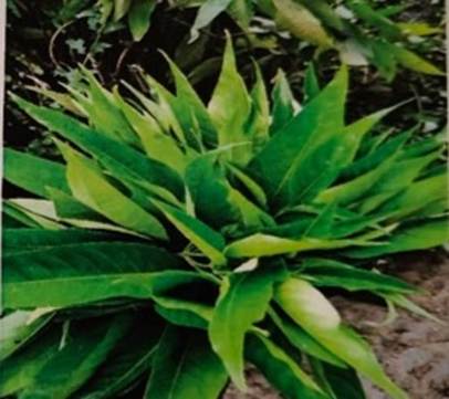
All chemicals and reagents used in the present study were of analytical grade. Fresh, healthy leaves of Adhatoda vasica were collected from Chandrana Halli village, Davanagere District, Karnataka, India ([Fig. 1]). The collected leaves were carefully cleaned under running tap water and then rinsed with distilled water to get rid of dust, soil particles, and other surface impurities. Later, the leaves were shade-dried at room temperature for four weeks until they reached a consistent weight. The dried leaves were subsequently ground into a fine powder using a mechanical grinder and stored in airtight containers at room temperature until further use for phytochemical screening and the green synthesis of zinc oxide nanoparticles (ZnO NPs)[19].
Fig. 1: Adhatoda vasica leaves used in the study
Synthesis of Zinc oxide nanoparticles from Adhatoda vasica leaves extract
Preparation of leaf extract:
The dried leaf powder of Adhatoda vasica leaves was used to prepare leaf extracts using different solvents including distilled water (aqueous), ethanol, and acetone[20].
Preparation of aqueous extract of Leaf: For the preparation of aqueous extract, the dried powder of Adhatoda vasica leaves were combined with deionised water in a 1:10 (w/v) ratio. The mixture was heated at 70°C and constantly stirred at 250 rpm using a magnetic stirrer for 30–45 minutes to enhance the extraction of bioactive phytoconstituents. In order to eliminate insoluble plant residues, the mixture was first allowed to cool to ambient temperature before being filtered through Whatman No. 41 filter paper. The filtrate was collected and stored at 4°C for the green production of zinc oxide nanoparticles (ZnO NPs) and for further phytochemical screening.
Preparation of Ethanol extract: To aid in the extraction of bioactive components, dried leaf powder of Adhatoda vasica (10 g) was extracted with 30 mL of 70% ethanol and incubated at 30°C for 12 hours on a rotary shaker. Following the initial extraction phase, 70% ethanol was added to the solvent volume to compensate for evaporative losses, and the extraction procedure was then continued for an additional twelve hours under identical conditions. To remove plant residues, the resulting mixture was filtered through Whatman No. 1 filter paper, and to eliminate any remaining particulate matter, the filtrate was centrifuged at 2000 rpm for 10 min. Then by solvent evaporation method, the supernatant obtained was concentrated to dryness. After that, the dried ethanolic extract was transferred to sterile containers and kept at 4°C until further use for biological activity and phytochemical screening.
Preparation of Acetone extract:For the preparation of acetone extract, 10 g of dried Adhatoda vasica leaf powder was combined with 45 mL of acetone and incubated at 30°C for 24 hours on a rotating shaker. To preserve the initial extraction volume, acetone was added back after extraction. After filtering the mixture through Whatman No. 1 filter paper, and to eliminate the residual particulate matter, the filtrate was centrifuged at 3000 rpm/10 minutes. Later, the supernatant was collected and concentrated by solvent evaporation method until it was completely dry. Before being used for phytochemical analysis and further analysis, the dried extract was stored in sterile containers and kept in a refrigerator at 4°C.
Qualitative phytochemical screening
Different solvent extracts (aqueous, ethanol, acetone) of Adhatoda vasica leaf were subjected to qualitative phytochemical screening to identify different bioactive secondary constituents by standard methods [19].
Test for alkaloids:
Hager's test: The plant extract was acidified with a few drops of dilute hydrochloric acid, then Hager's reagent (aqueous solution of picric acid) was added. Formation of yellow precipitate indicated the presence of alkaloids.
Mayer's test:Mayer's reagent (mercuric chloride-1.36 g, potassium iodide-5 g dissolved in 100 ml of distilled water) was added to the leaf extract and observed for white or green colour precipitate which indicated the presence of alkaloids.
Test for reducing sugars:
Benedict's test: To 1 ml of plant extract, 2 ml Benedict's reagent was added, heat the mixture in a boiling water bath for 2 minutes and allowed it to cool. Orange or brick red precipitate indicated the presence of reducing sugars.
Fehling's test:To 1 ml of plant extract, 2 ml of Fehling's reagent (equally mixed Fehling's solution A and B) was added, then heated in boiling water for a few minutes and allowed to cool. Formation of brick-red precipitate indicated the presence of reducing sugars.
Test for glycosides:
The plant extract was mixed with a few drops of diluted sulfuric acid and 20% KOH solution. To this, 3 ml of Fehling's reagent was added and heated in water bath for few minutes. The presence of glycosides was indicated by the appearance of brick red colour.
Test for phenols:
Ferric chloride test:To 2 ml of plant extract, few drops of 5% ferric chloride (FeCl₃) solution was added. Bluish black or dark colouration confirms the presence of phenols.
Test for flavonoids:
Alkaline reagent test:A few drops of 10% sodium hydroxide solution was added to 2 ml of plant extract and the colour change was observed, then dilute hydrochloric acid (HCl) was added. Formation of intense yellow colour on addition of HCl indicated the presence of flavonoids.
Test for terpenoids:
Salkowski test:2 ml of chloroform was added to 2 ml of plant extract in a test tube and concentrated sulfuric acid was added carefully and the development of reddish-brown or golden-yellow coloration at the interface confirms the presence of terpenoids.
Test for steroids:
Liebermann-Burchard test:1–2 ml of plant extract was dissolved in 2 ml of chloroform with 2 ml of acetic anhydride. Concentrated sulfuric acid was added and the formation of emerald green or blue-green colour indicates the presence of steroids.
Test for tannins:
To 1–2 ml of plant extract, two to three drops of 5% ferric chloride solution were added and observed for blue-black or greenish-black colour formation that indicates the presence of tannins.
Characterisation of green synthesised Zinc oxide (ZnO) nanoparticles
FTIR analysis:
Fourier transform infrared spectroscopy (FTIR) Bruker ALPHA II spectrophotometer was used to identify the functional groups responsible for the reduction, stabilisation, and capping of the synthesized nanoparticles from Adhatoda vasica leaves. Resulting spectra were recorded in the mid-infrared region spanning from 400-4000 cm-1 at a resolution of 4 cm-1[21].
XRD analysis:
The crystalline nature and phase composition of the synthesised nanoparticles were identified using X-ray diffraction (XRD). The dried nanoparticles were finely milled and then placed on a sample holder for analysis. XRD patterns were recorded using an X-ray diffractometer operating at 40 kV and 30 mA with Cu-Kα radiation (λ = 1.5406 Å). The diffraction data were collected across a 2θ range of 5–80° using a scanning rate of 2° min⁻¹ and a step size of 0.02°. The crystalline structure and purity of the produced nanoparticles were verified by additional analysis of the diffraction pattern[22].
SEM analysis:
Scanning electron microscopy (SEM) was used to analyse the microstructural and surface morphology features of the produced nanoparticles. Double-sided conductive carbon tape was used to attach the powdered dry nanoparticles to an aluminium stub coated with carbon. Before analysis, the samples were sputter-coated with a small layer of gold to increase conductivity and reduce charging effects during imaging. High vacuum conditions and an accelerating voltage of 5–20 kV were used for SEM studies. To assess the distribution, degree of aggregation, surface texture, and particle shape, micrographs were taken at various magnifications[23].
Antibacterial activity of Adhatoda vasica leaf extracts and ZnO nanoparticles
The antibacterial activity of Adhatoda vasica leaf extracts and synthesized nanoparticles was evaluated against test pathogens that include Staphylococcus aureus, Escherichia coli, coagulase-negative Staphylococcus, Klebsiella pneumoniae, and Candida sp using the agar well diffusion method. Autoclaved Muller-Hinton agar (MHA) was poured into petri plates. The test pathogens were seed inoculated and wells of 6 mm were made with a sterile cork borer. Leaf extracts prepared with aqueous, acetone and ethanol solvents were added to the wells at a concentration of 50 mg ml-1 and nanoparticles at concentrations of 60 mg ml-1, 40 mg ml-1and 20 mg ml-1were added to separate wells; the plates were incubated at 37°C for 24 hours and antibacterial activity was assessed by measuring the diameter of the zone of inhibition (ZOI) and recorded in millimeters (mm)[24] and DMSO was used as control.
RESULTSQualitative phytochemical screening
The results of phytochemical screening of Adhatoda vasica leaf extract prepared from different solvent extracts showed that the plant is rich in various bioactive secondary metabolites such as alkaloids, flavonoids, tannins, phenolic compounds, glycosides and steroids. This suggests the potential antibacterial and therapeutic properties of Adhatoda vasica as these compounds possess anti-inflammatory, antioxidant and other pharmacological activities.
[Table. 1] displays the findings of the qualitative phytochemical screening of Adhatoda vasica leaf extracts made with ethanol, acetone, and aqueous solvents. These findings showed that the phytochemical content differed between the various solvent extracts, suggesting variations in the solvents' extraction consequently their effectiveness.
Sl. No.
Phytochemical tests
Aqueous
Acetone
Ethanol
1
Alkaloids test
+
+
+
2
Reducing sugar
+
+
+
3
Glycosides
+
-
-
4
Phenolic compounds
+
+
+
5
Flavonoids
+
+
+
6
Terpenoids
+
+
-
7
Phytosterols
-
-
-
8
Tannins
+
+
+
Table 1: Qualitative phytochemical screening of aqueous, acetone and ethanolic leaf extracts of Adhatoda vasica. The presence (+) or absence (-) of major phytochemical constituents was determined using standard qualitative phytochemical tests
(+) presence ofphytochemical constituents
(-) absence phytochemical constituents
Alkaloids were found in all three extracts, indicating that these nitrogen-containing secondary metabolites are extensively distributed in A. vasica leaves. Reducing sugars were also found in the ethanolic, acetone, and aqueous extracts, indicating that these compounds can be extracted from both polar and moderately polar solvents.
All three extracts also included flavonoids and phenolic substances. These findings showed that A. vasica leaves are rich in polyphenolic components, which may be efficiently extracted using ethanol, acetone, and aqueous solvents, indicating that these substances are important bioactive components of the plant.
Glycosides, on the other hand, were absent in acetone and ethanolic extracts and only found in the aqueous extract. This result implies that glycosidic chemicals were more successfully extracted by water, most likely because of their greater polarity and solubility in aqueous conditions.
The ethanolic extract did not contain terpenoids, while it was present in aqueous and acetone extracts. Differences in solvent polarity and the chemical properties of these chemicals may be the cause of the terpenoids' varying occurrence among the extracts.
Tannins were present in all the solvent extracts. None of the solvent extracts included phytosterols, suggesting that they were either absent or present at levels below the qualitative assays' detection limits.
These results showed that the qualitative phytochemical profile of A. vasica sample was significantly influenced by the extraction solvent selection. Alkaloids, phenolic compounds, flavonoids, glycosides, and terpenoids are examples of bioactive secondary metabolites that are abundant in A. vasica and may be responsible for its purported pharmacological and antibacterial effects.
Green synthesis of zinc oxide nanoparticles using Adhatoda vasica leaves extract
The green synthesis of zinc oxide nanoparticles using Adhatoda vasica leaf extract was confirmed by a discernible colour shift during the reaction. The aqueous leaf extract was initially light green in colour, whereas the zinc sulfate solution was colourless. When the leaf extract and zinc sulfate solution were combined, the reaction mixture gradually changed from light green to dark brown, indicating the formation of ZnO nanoparticles. The characteristic colour change is caused by bioactive phytochemicals in the leaf extract that transform Zn²⁺ ions into ZnO nanoparticles. The production of nanoparticles was evident throughout the incubation period, suggesting that Adhatoda vasica's biomolecules effectively served as stabilising and reducing agents during the nanoparticle synthesis process.
The effective biogenesis of ZnO nanoparticles was further verified by physicochemical characterisation using Fourier Transform Infrared Spectroscopy (FTIR), X-ray Diffraction (XRD), and Scanning Electron Microscopy (SEM). These methods verified the synthesised nanoparticles' optical characteristics, surface morphology, crystalline structure, and functional groups involved in nanoparticle stabilisation.
Characterisation of green synthesised Zinc oxide (ZnO) nanoparticles
FTIR:
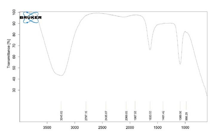
FTIR spectrometry was used to determine the nature of functional groups existing in the ZnO NPs synthesized from Adhatoda vasica leaf extract ([Fig. 2]). The FTIR of the synthesized ZnO nanoparticles was performed in the mid-infrared region in the range of 500 to 3500 cm⁻¹ and revealed some absorption bands which indicate characteristic bioactive functional groups from the plant extract.
The broad absorption band at 3273.71 cm⁻¹ indicates O-H stretching vibrations, and the existence of hydroxyl groups and hydrogen bonding phenols that are crucial in the reduction and stabilization of nanoparticles. The absorption band at 3134.06 cm⁻¹ indicates N-H stretching or aromatic C-H stretching vibrations and indicates the presence of amine or aromatic phytochemicals. The absorption peaks at 3024.58 cm⁻¹ indicate aliphatic C-H stretching vibrations, showing the existence of hydrocarbon groups that could be from biomolecules in the plant extract. A distinct absorption spectrum at 1826.16 cm⁻¹ is due to stretching vibration of carbonyl (C=O) group and is possibly due to organic acids or ester groups used in nanoparticle synthesis. The major absorption peak at 1621.71 cm⁻¹ is due to C=C stretching vibrations of aromatic rings or amide band. The absorption spectrum at 1062.73 cm⁻¹ is for C-O stretching vibrations of alcohols, ethers or esters. The absorption peak at 870.25 cm⁻¹ is for aromatic C-H out-of-plane bending vibrations. Thus, FTIR analysis indicates that the phytochemicals from Adhatoda vasica leaves extract have participated in the reduction of Zn²⁺ ions and also in capping and stabilisation of the synthesized ZnO nanoparticles.
Fig. 2: FTIR spectrum of biosynthesized zinc oxide nanoparticles synthesized using Adathoda vasica leaf extract. The spectrum shows the absorption bands corresponding to functional groups of phytochemicals involved in the stabilisation, capping and reduction of ZnO nanoparticles
XRD:
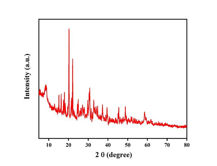
The effective production of crystalline zinc oxide was verified by the X-ray diffraction (XRD) pattern of the biosynthesised ZnO nanoparticles are depicted in [Fig. 3]. X-ray diffraction (XRD) examination verified the successful synthesis of crystalline zinc oxide nanoparticles. The crystalline structure of the biosynthesized ZnO nanoparticles was demonstrated by the diffraction pattern, which showed multiple distinct and sharp peaks. The observed diffraction peaks are consistent with the standard JCPDS data (Card No. 36-1451) and are typical of the hexagonal wurtzite crystal structure of ZnO. The absence of notable impurity peaks showed great phase purity, while the high intensity and narrow diffraction peaks show good crystallinity of the produced nanoparticles. Remaining biomolecules from the biological extract, which function as reducing and capping agents during the creation of nanoparticles, may be responsible for the broad background seen at lower diffraction angles. Overall, the successful biosynthesis of crystalline ZnO nanoparticles was confirmed by the XRD data.
Fig. 3: X-ray diffraction patterns of biosynthesized ZnO nanoparticles showing distinct diffraction peaks, confirming the crystalline nature and successful formation of ZnO nanoparticles
SEM analysis:
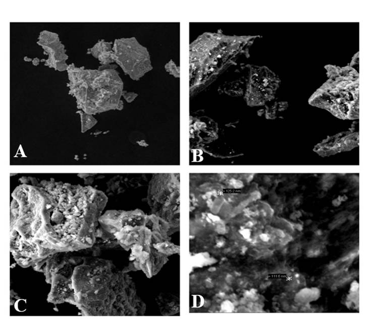
The surface shape and particle size of the biosynthesised nanoparticles were examined using scanning electron microscopy (SEM); micrographs are displayed in [Fig. 4].
Fig. 4: SEM micrographs of biosynthesised ZnO nanoparticles synthesized with Adhatoda vasica leaf extract at various magnifications: (A) 500×, (B) 1,000×, (C) 2,500×, and (D) high-magnification image displaying ZnO nanoparticles with particle sizes ranging from roughly 111.6 to 125.2 nm
Figure A: The low-magnification SEM image shows the overall shape of the generated nanoparticles. The particles appear as aggregates of varying sizes that resemble plates or flakes. Particle aggregation may be caused by the nanoparticles' increased surface energy during the drying process.
Figure B: At a higher magnification, the nanoparticles' surface was rough and uneven, with irregular edges. The particles remained aggregated, but the individual crystalline domains became more visible. This type of aggregation is commonly observed in biosynthesised nanoparticles because phytochemicals serve as capping and reducing agents.
Figure C: Further imaging reveals compact aggregates consisting of many small nanoparticles scattered over the particle surface. The surface's porous and uneven look showed that crystalline nanoparticle clusters had formed.
Figure D: The highest magnification SEM image clearly displayed nanosized particles with an average particle size ranging from approximately 111 to 125 nm, according to the scale measurements on the micrograph. The irregular form and slight aggregation of plant-mediated synthesised nanoparticles are their defining characteristics.
The SEM analysis confirmed the successful production of nanoparticles with distinctive nanoscale morphology. Most of the particles were irregular to quasi-spherical in shape and had rough surfaces. Furthermore, the SEM micrographs revealed that the nanoparticles were densely packed aggregates. This aggregation, commonly observed in green-synthesised nanoparticles, may be caused by the high surface energy of the nanoparticles and the interaction of the phytochemicals in the leaf extract of Adhatoda vasica which function as reducing and capping agents.
Antibacterial activity of Adhatoda vasica leaf extracts and ZnO nanoparticles
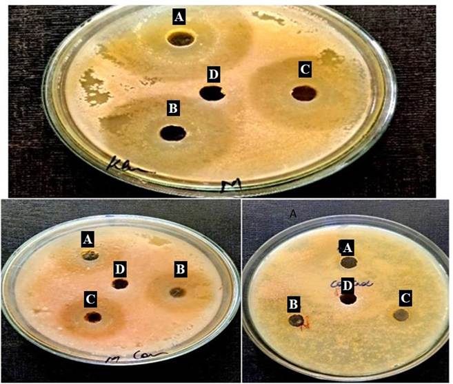
The agar well diffusion assay was used to assess the antibacterial activity of Adhatoda vasica leaf extracts and biosynthesised ZnO nanoparticles against Staphylococcus aureus, Escherichia coli, coagulase negative Staphylococcus aureus (CNSA), Klebsiella pneumoniae, and Candida sp, was measured as Zone of inhibition (ZOI).For A. vasica leaf extract (aqueous) showed maximum ZOI against CNSA and K. pneumoniae (15 ± 0.36 mm and 15 ± 0.41mm) while acetone extract was inhibiting only E. coli (ZOI 11±0.32 mm). However, ethanol extract was consistently inhibiting all four bacterial isolates with maximum ZOI against K. pneumoniae but Candida sppwas resistant ([Fig. 5] and [Table. 2]).
The antibacterial activity of the biosynthesised ZnO nanoparticles was concentration-dependent. At different concentration of nanoparticle,
Fig. 5:The antibacterial efficacy of the different concentrations of ZnO nanoparticles A: 60 mg/ml, B: 40mg/ml, C: 20 mg/ml and D: DMSO (control) against test pathogens 1. Klebsiella pneumoniae, 2. Candida spp, and 3. Various solvent extracts A: aqueous, B: ethanol, and C: acetone by agar well diffusion method against Klebsiella pneumoniae. The control DMSO is represented by C. Antibacterial effectiveness is indicated by zones of inhibition (in mm)
ZOI was found to be maximum at 60 mg mL⁻¹ with 17 ± 0.63 mm, 11 ± 0.39 mm, and 39 ± 0.63 mm against Escherichia coli, CNSA, Klebsiella pneumoniae respectively while no activity wasobserved against S. aureus but Candida sp. showed 20 ± 0.39 mm of ZOI ([Table. 2]). Klebsiella pneumoniae exhibited maximum ZOI, with range of ZnO NP concentration with decrease in ZOI with decrease in NP concentration.However, when compared to the other tested microbial species for ZnO NP antimicrobial activity, Klebsiella pneumoniae was maximum with its ZOI with all NP concentration (39 ± 0.63 mm, 37 ± 0.83 mm, and 34 ± 0.71 mm for 60, 40 and 20 mg mL⁻¹). Furthermore, the ZnO NP demonstrated notable antifungal efficacy against Candida sp. with ZOI of 20 ± 0.96 mm, 23 ± 0.54 mm, and 28 ± 0.66 mm (60, 40 and 20 mg mL⁻¹ respectively), an increase in ZOI with decrease in NP concentration was a notable point.
In total, ethanolic and acetone leaf extracts showed observable antibacterial activity, but maximum ZOI was observed with aqueous leaf extract. When compared to the leaf extracts, the biosynthesised ZnO nanoparticles showed stronger antibacterial action, especially against Klebsiella pneumoniae along with other tested organisms. These results imply that the ZnO NP are potential with improved the antimicrobial activity when compared to leaf extract of Adhatoda vasica.
Extracts and Nanoparticles
Concentration, mg/ml
Zone of inhibition (ZOI) against different test pathogens measured in mm
Staphylococcus aureus
Escherichia coli
Coagulase-negative Staphylococcus
Klebsiella pneumoniae
Candida spp.
Leaf extracts
a. Aqueous
50
14±0.83
13±0.45
15±0.36
15±0.41
-
b. Acetone
50
-
11±0.32
-
-
-
c. Ethanol
50
12±0.22
12±0.51
12±0.76
15±0.38
-
Nanoparticles
60
-
17±0.63
11±0.39
39±0.63
20±0.96
40
-
16±0.47
12±0.99
37±0.83
23±0.54
20
-
15±0.38
15±0.56
34±0.71
28±0.66
Table 2: The antibacterial activity of Adhatoda vasica leaf extracts (aqueous, acetone, and ethanol) and biosynthesised ZnO nanoparticles against bacterial and fungal pathogens evaluated using the agar well diffusion assay
The data are expressed as the mean zone of inhibition (mm) ± standard deviation (SD).
(-) = No inhibition observed; values are expressed as mean ± SD (n=3)
DISCUSSION
The present study investigated the phytochemical profile of Adhatoda vasica leaf extracts prepared in three solvents (aqueous, acetone and ethanol). The antibacterial efficacy of both the crude extracts and the biosynthesised nanoparticles (ZnO NPs) against clinically important pathogens was evaluated. The results of the current study corroborate with previous findings about Adhatoda vasica and emphasise its viability as a sustainable biological resource for the production of ZnO nanoparticles with increased antibacterial activity. In the current investigation, alkaloids, reducing sugars, glycosides (in the aqueous extract only), phenolic compounds, flavonoids, tannins and terpenoids were found in A. vasica leaf extracts using qualitative phytochemical screening while phytosterols were not found. These findings are in consistent with previous phytochemical analyses of the plant. Gohel, et al.[25] revealed the importance of these secondary metabolites for the biological activities of the plant and obtained positive results for alkaloids, steroids, saponins, phenols, and terpenoids in ethanolic leaf extracts of A. vasica. Similarly, Khan, et al.[26] validated the presence of alkaloids, flavonoids, terpenoids, tannins, and glycosides in the phytochemical analysis of A. vasica produced from Khyber Pakhtunkhwa. Alkaloids, phenolic compounds, and flavonoids were consistently found in all three solvent extracts in our investigation, supporting findings by
Sharma, et al.[29] that the biological activity of A. vasica is mostly dependent on phenolic group secondary metabolites. The solvent-dependent variation was well observed in this study, especially the limitation of glycosides to the aqueous extract and the lack of terpenoids in ethanol, is consistent with previous findings by Vasika, et al.[27], in which the polarity of the solvent determines extraction efficiency and, consequently, the phytochemical profile obtained from A. vasica leaves.
Based on the antibacterial activity of A. vasica leaf extracts performed by agar well diffusion method at 50 mg mL⁻¹ concentration, the aqueous extract was the most successful in suppressing the pathogens tested for S. aureus, E. coli, coagulase-negative Staphylococcus and K. pneumoniae. The ethanolic extract likewise showed a broad spectrum inhibitory effect (12–15 mm), despite the acetone extract's mild inhibition (for E. coli only 11 ± 0.32 mm) indicating the potential of A. vasica leaf extract. These results are consistent with the work conducted by Josephin, et al.[27]where A. vasica extract exhibited dose-dependent antibacterial activity, particularly against Gram-positive isolates, in ethanol, acetone, ethyl acetate, and petroleum ether.
Similarly, Gohel, et al.[25] found that ethanolic leaf extracts had the largest inhibitory zones against Aspergillus clavatus and Pseudomonas aeruginosa. Mishra, et al.[28] found that methanolic A. vasica extracts evaluated at 25 µg mL⁻¹ had broad-spectrum antibacterial activity against K. pneumoniae, E. coli, and S. aureus. The effectiveness of water as an eco-friendly extraction medium for polar phytoconstituents like alkaloids and flavonoids was demonstrated by the aqueous extract was used in the current study, which had comparable or better inhibition zone diameters against the same genera at the standardised concentrations.
The colour shift from light green to dark brown, which signified the reduction of Zn²⁺ ions into ZnO nanoparticles, confirmed the green chemistry synthesis of ZnO nanoparticles employing A. vasica aqueous leaf extract. The characterisation results (FTIR, XRD, and SEM) unequivocally showed that the products obtained in the study were ZnO nanoparticles. Another highly pertinent example by Pandian, et al.[30], their research group obtained phase-pure crystalline ZnO NPs with a very small crystallite size of 5.2 nm using Justicia adhatoda (syn. A. vasica) aqueous leaf extract as both the reducing and stabilising agent. Additionally, this study demonstrated the improved antibacterial performance of biologically generated ZnO NPs, demonstrating the efficacy of A. vasica phytochemicals as a stabilising, reducing, and capping agent.
The employment of phytochemicals in the biosynthesis of nanoparticles was indicated by prominent peaks seen in the FTIR analysis of the biosynthesised ZnO NPs in this study. The existence of hydrogen bonds and O-H groups, which are typical of phenolic phytochemicals utilised as primary reducing agents, is shown by the broad peak of roughly 3273 cm⁻¹. The peaks match phenols, proteins, and carboxylate groups as reducing agents and capping molecules[34] was also demonstrated in Croton macrostachyus. Using the same Bruker Alpha II FTIR spectrometer, Santhosh, et al.[35] found identical functional groups (O-H, N-O, C-O, and C=C) in ZnO NPs produced from Catharanthus roseus. The presence of O-H, C=O, C=C, and C-O functional groups in the present ZnO NPs suggests that synthesis, stabilisation, and capping of the nanoparticles were significantly influenced by the phytochemical compounds in A. vasica, particularly the alkaloids, phenols, and flavonoids, consistent[33].
Through distinct diffraction peaks of crystalline ZnO and the absence of any extra peaks caused by contaminants, XRD examination verified the crystalline nature of the biosynthesis of ZnO NPs. The findings are in line with earlier research that consistently confirmed the wurtzite hexagonal crystal structure of ZnO NPs made from plants. For example, Suresh, et al.[31] synthesised ZnO NPs using Cinnamomum verum bark extract and demonstrated their crystalline nature via XRD examination. Using J. adhatoda plant extract, Pandian, et al.[30] produced a very small crystallite size of 5.2 nm, demonstrating the impact of phytochemical content on the crystal development process.
ZnO nanoparticles with a size range of 111 to 125 nm were successfully formed, as per SEM measurements. It was demonstrated that the nanoparticles had a rough surface, an irregular to quasi-spherical morphology, and some degree of aggregation—all of which are expected outcomes of employing plants to make nanoparticles. These nanoparticle properties are comparable to those discovered in previous research on green synthesised ZnO nanoparticles[30, 36-38].
The biogenic ZnO nanoparticles exhibited intense antibacterial activity against the investigated microorganisms in a dose-dependent manner when compared to the raw leaf extract of Adhatoda vasica with ZOI of 39 ± 0.63 mm, 37 ± 0.83 mm, and 34 ± 0.71 mm at 60, 40, and 20 mg mL⁻¹, respectively, the highest antibacterial activity was observed against Klebsiella pneumoniae. The combination of several processes, including the production of reactive oxygen species (ROS), the release of zinc ions, the rupture of cell membranes, and the bioactive phytochemicals of A. vasica that stayed affixed to the nanoparticle's surface, can be responsible for the increased antibacterial activity. Furthermore, none of the raw extracts exhibited any antifungal activity, whereas the ZnO nanoparticles showed strong antifungal activity against Candida spp demonstrating the potential of biogenic ZnO NP. Moreover, ZnO NP of A. vasica showed highest ZOI (28 ± 0.66 mm) at lowest concentration (20 mg/ml) indicating an inverse dose response relationship. This may be due to, for higher concentration of ZnO NP, Candida spp would have become resistant gradually or sublethal dosage would have triggered the organism to develop adaptive ZnO NP stress response leading to increased growth potential of the tested unicellular fungi.
Numerous mechanisms, including the production of ROS, the release of Zn²⁺ ions, and membrane contact, could account for the enhanced antibacterial activity of zinc oxide nanoparticles[32]. In present study biogenic ZnO NP's small size results in a large surface area, which increases the rate at which they interact with microbial cells and enhance their antibacterial activity. This study's concentration-dependent antibacterial activity is consistent with previous findings of ZnO nanoparticles derived from other plants which re-authenticate the usage of A. vesica for clinical purpose.
CONCLUSION
The current study effectively produced ZnO nanoparticles by green synthesis using Adhatoda vasica, along withaqueous, ethanolic, and acetone extracts from the leaves which also served as reducing and stabilising agents. Qualitative phytochemical screening was used to identify secondary metabolites such as alkaloids, flavonoids, phenolics, tannins, saponins, and terpenoids that facilitated the bioreduction and stabilisation of ZnO nanoparticles. The synthesised nanoparticles were characterised using FTIR, XRD, and SEM techniques. FTIR analysis showed the role of phytoconstituents in the creation of nanoparticles, XRD analysis revealed the development of highly crystalline zinc oxide nanoparticles with a hexagonal wurtzite crystal structure, while SEM study revealed nanoparticles with various surface morphologies.
Since the biologically produced ZnO nanoparticles were found to have higher antibacterial potential against both Gram-positive and Gram-negative bacteria when compared to the equivalent leaf extracts, indicating the potential of ZnO nanoparticles. ZnO nanoparticles with exceptional antibacterial and anti-Candida spp qualities overweighs with respect to future clinical use.
In conclusion, the results showed that ZnO nanoparticles produced by Adhatoda vasica are economical, sustainable, and possess potent antimicrobial properties. These biological nanoparticles have potential applications in pharmacology, medicine, and antibacterial therapy. To better understand their cytotoxicity, biocompatibility, antibacterial mechanism, and in vivo efficacy, advanced study is necessary.
DISCLOSURE
Funding:
The authors gratefully acknowledge the financial support provided by KSTePS, DST, Government of Karnataka (Karnataka Science and Technology Promotion Society, Department of Science and Technology), Vision Group on Science and Technology (VGST), for sponsoring this project (VGST/GRD-1017/CISEE).
Author's Contribution:
SMP executed the experiment, GD conceived the idea and supervised the project, SR analysed data, SGS and NM were involved in the conception and design of the study, participated in collection of data, performed the experiments, VS analysed data. All authors have read and approved the final manuscript.
Ethics Approval and Consent to Participate:
Not applicable. The present study did not involve human participants, human tissues, clinical samples, or animals.
Patient Consent for Publication:
Not applicable.
Competing Interests:
The authors declare that they have no competing interests.
References
Cowan MM
Plant Products as Antimicrobial AgentsClinical Microbiology Reviews1999124https://doi.org/10.1128/cmr.12.4.564
Rates SMK
Plants as source of drugsToxicon2001395https://doi.org/10.1016/s0041-0101(00)00154-9
Cushnie TPT, Lamb AJ
Antimicrobial activity of flavonoidsInternational Journal of Antimicrobial Agents2005262https://doi.org/10.1016/j.ijantimicag.2005.09.002
Ventola CL
The antibiotic resistance crisis. Part 1: Causes and ThreatsP T2015404https://pmc.ncbi.nlm.nih.gov/articles/PMC4378521/
Daglia M
Polyphenols as antimicrobial agentsCurrent Opinion in Biotechnology2012232https://doi.org/10.1016/j.copbio.2011.08.007
Silva NCC, Fernandes Jr A
Biological properties of medicinal plants: a review of their antimicrobial activityJournal of Venomous Animals and Toxins including Tropical Diseases2010163https://doi.org/10.1590/s1678-91992010000300006
Subapriya R, Nagini S
Medicinal Properties of Neem Leaves: A ReviewCurrent Medicinal Chemistry-Anti-Cancer Agents200552https://doi.org/10.2174/1568011053174828
Claeson UP, Malmfors T, Wikman G, Bruhn JG
<I>Adhatoda vasica</I>: A critical review of ethnopharmacological and toxicological dataJournal of Ethnopharmacology2000721-2https://doi.org/10.1016/s0378-8741(00)00225-7
Dhankhar S, Kaur R, Ruhil S, Balhara M, Dhankhar S, Chhillar AK
A review on <I>Justicia adhatoda</I>: A potential source of natural medicineAfrican Journal of Plant Science2011511https://academicjournals.org/journal/AJPS/article-full-text-pdf/EFA667010942
Iravani S
Green synthesis of metal nanoparticles using plantsGreen Chemistry20111310https://doi.org/10.1039/c1gc15386b
Singh J, Dutta T, Kim KH, Rawat M, Samddar P, Kumar P
‘Green’ synthesis of metals and their oxide nanoparticles: applications for environmental remediationJournal of Nanobiotechnology2018161https://doi.org/10.1186/s12951-018-0408-4
Raghupathi KR, Koodali RT, Manna AC
Size-Dependent Bacterial Growth Inhibition and Mechanism of Antibacterial Activity of Zinc Oxide NanoparticlesLangmuir2011277https://doi.org/10.1021/la104825u
Jones N, Ray B, Ranjit KT, Manna AC
Antibacterial activity of ZnO nanoparticle suspensions on a broad spectrum of microorganismsFEMS Microbiology Letters20082791https://doi.org/10.1111/j.1574-6968.2007.01012.x
Sirelkhatim A, Mahmud S, Seeni A, Kaus NHM, Ann LC, Bakhori SKM, <I>et al</I>
Review on Zinc Oxide Nanoparticles: Antibacterial Activity and Toxicity MechanismNano-Micro Letters201573https://doi.org/10.1007/s40820-015-0040-x
Padmavathy N, Vijayaraghavan R
Enhanced bioactivity of ZnO nanoparticles—an antimicrobial studyScience and Technology of Advanced Materials200893https://doi.org/10.1088/1468-6996/9/3/035004
Harborne JB
<I>Phytochemical Methods. A Guide to Modern Techniques of Plant Analysis</I>. London: Chapman and Hall Ltd.1973https://archive.org/details/phytochemicalmet0000harb
Pachaiappan R, Rajendran S, Kumar PS, Vo D-VN, Hoang TKA
Green Synthesis of Zinc Oxide Nanoparticles by <I>Justicia adhatoda</I> Leaves and Their Antimicrobial ActivityChemical Engineering & Technology2021443https://doi.org/10.1002/ceat.202000470
Mahajan M, Kumar S, Gaur J, Kaushal S, Dalal J, Singh G, <I>et al</I>
Green synthesis of ZnO nanoparticles using
<i>Justicia adhatoda</i>
for photocatalytic degradation of malachite green and reduction of 4-nitrophenolRSC Advances2025154https://doi.org/10.1039/d4ra08632e
Monshi A, Foroughi MR, Monshi MR
Modified Scherrer Equation to Estimate More Accurately Nano-Crystallite Size Using XRDWorld Journal of Nano Science and Engineering20120203https://doi.org/10.4236/wjnse.2012.23020
Roy VC, Johnwilmet PL, Anabalagan R, Krishnan J, Balakrishnan AS
Characterization and Evaluation of <I>Adhatoda vasica</I> and its green synthesized Nanoparticles for its efficacy in <I>Culex quinquefasciatus</I>2025https://doi.org/10.21203/rs.3.rs-7822392/v1
Latha M, Priyanka M, Rajasekar P, Manikandan R, Prabhu NM
Biocompatibility and antibacterial activity of the <I>Adhatoda vasica</I> Linn extract mediated silver nanoparticlesMicrobial Pathogenesis201693https://doi.org/10.1016/j.micpath.2016.01.013
Gohel A, Upadhye V, Upadhyay TK, Rami E, Panchal R, Jadhav S, <I>et al</I>
Study on Phytochemical Screening and Antimicrobial Activity of <I>Adhatoda vasica</I>Canadian Journal of Medicine202133https://doi.org/10.33844/cjm.2021.60509
Khan SI, Ali A, Ahmad N, <I>et al</I>
Phytochemical analysis and antimicrobial activity of <I>Adhatoda vasica</I> leavesPure and Applied Biology202092https://doi.org/10.19045/bspab.2020.90174
Suresh J, Yuvakkumar R, Nathanael AJ, Hong SI, Rajendran V
Green synthesis of ZnO nanoparticles using <I>Cinnamomum verum</I> bark extract and their antibacterial potentialityCatalysts2020https://doi.org/10.3390/catal10030335
Siddiqui MA, Ahmad J, Farshori NN, <I>et al</I>
Green synthesis of zinc oxide nanoparticles using aqueous fruit extracts of <I>Myristica fragrans</I>: characterizations and biological and environmental applicationsACS Omega2021614https://doi.org/10.1021/acsomega.1c00310
Kirana P, Prashantkumar CS, Soundarya R, Shwetha GS, Kalasad MN, Gayathri D<I>et al</I>
<I>Tinospora cordifolia's</I> Green Synthesis Approach: Comparative Study on Biogenic and Chemical Synthesis of Zinc Oxide and Zinc Sulfide NanoparticlesMaterials International202464https://doi.org/10.33263/materials64.040
Chakra PS, Banakar, A, Puranik SN, Kaveeshwar V, Ravikumar CR, Gayathri D
Characterization of ZnO nanoparticles synthesized using probiotic <I>Lactiplantibacillus plantarum</I> GP258Beilstein Journal of Nanotechnology202516https://doi.org/10.3762/bjnano.16.8
Sharath SC, Gayathri D, Santosh RM, Kalasad MN
Growth and characterization of Ag2S semiconductor nanoparticlesMaterials Today: Proceedings202267https://doi.org/10.1016/j.matpr.2022.08.159